Cilia and Flagella
Cilia and flagella are two cell organelles primarily found in protozoa, such as amoeba, paramecium, and euglena. However, they are also found in other microorganisms, such as bacteria, algae, and fungi, and in animals, except for higher plants.
They are motile cellular appendages made of microtubules and remain covered by the plasma membrane. Both cilia and flagella aid in movement.
Compare and Contrast Cilia and Flagella
Although cilia and flagella share many characteristics, they are also different in a number of ways.
Difference between Cilia and Flagella
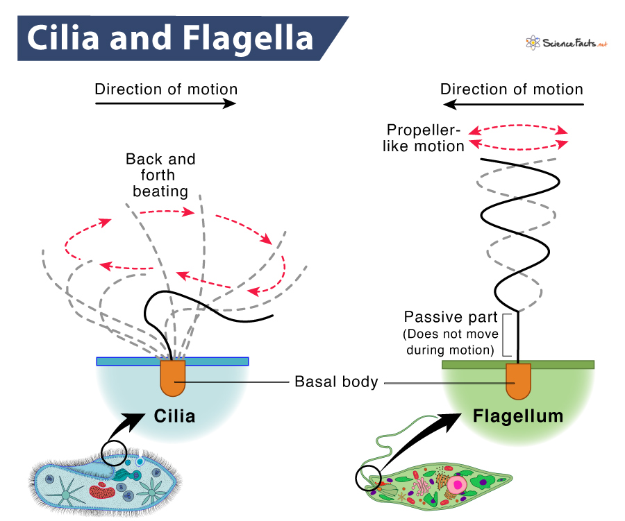
Cilia are short, microscopic, hair-like structures or organelles extending in large numbers from the surface of eukaryotic cells. In contrast, flagella are long, complex filamentous cytoplasmic structures protruding through the cell wall.
| Basis | Cilia | Flagella |
|---|---|---|
| 1. Found | Only in eukaryotes such as areas of the respiratory tract and female reproductive track | Both eukaryotes and prokaryotes |
| 2. Composed Of | Polymers of globular proteins called tubulin, the motor protein called dynein, and nexin | Polymers of tubulin and dynein. Bacterial flagella also contain flagellin |
| 3. Structure and Organization | Length: Short, hair-like, (5-10)µ in length Thickness: Around 0.3 to 0.5 μm in thickness Organization: Possess a central bundle of microtubules called the axoneme 9 outer doublet microtubules surround a central pair of singlet microtubules When viewed under an electron microscope, the cross-section of the axoneme shows the characteristic 9 + 2 arrangement | Length: Long, whip-like, or thread-like organelle. 150µ in length Thickness: Around 0.02 to 0.025 μm in thickness Organization: Eukaryotic flagella are remarkably similar in their organization to cilia, showing the characteristic 9 + 2 arrangement of microtubules Prokaryotic flagella are simpler structures made up of flagellin |
| 4. Functions | Helps primarily in locomotion. It also helps in feeding circulation, aeration, and respiration | Help mainly in locomotion |
| 5. Beating Pattern | Complicated, can move in a wide range of motions | Circular, wave-like, or propeller-like motion |
| 6. Distribution in Cell | Distributed throughout the cell surface | Present at one end or two ends or all over the surface |
| 7. Density | Numerous, approximately hundreds per cell | Fewer than cilia, less than 10 per cell |
| 8. Energy Requirement | Use ‘kinesin,’ having an ATPase activity that produces energy to perform the movement | Powered by the proton-motive force generated in the plasma membrane in prokaryotes. It is ATP-driven in eukaryotes |
| 9. Position In Cell | Present throughout the surface of the cell | Either present at both the ends of the cell or all over the surface |
| 10. Types | Two types: motile or non-motile | Three types: bacterial, archaeal, and eukaryotic |
| 11. Types of Motion | a) Rotational, like a motor b) Very fast-moving | a) Wave-like, sinusoidal undulating movement b) Slow moving |
| 12. Beating Pattern | Beating in a coordinated rhythm either simultaneously (synchronous) or one after the other (metachronous) | Beating is independent of each other |
| 13. Cross section | Nexin arm present | Nexin arm absent |
How are Cilia and Flagella Alike
Although they differ in many aspects, cilia and flagella have similarities. They are
- Function primarily in locomotion.
- Arise from the basal body, a centriole-like structure
- Develop from the cell’s plasma membrane
- Have an axoneme, a central filament. Axoneme has 11 microtubules, of which 9 are in pairs (doublet), and the remaining 2 are located in the center (singlet)., showing the typical 9+2 arrangement of microtubules
Conclusion
Cilia and flagella are two significant microscopic appendages of cells. Cilia are present in both animals and microorganisms but absent in higher plants. In contrast, flagella are used for mobility in bacteria and gametes (sperm and egg cells) of eukaryotes. Both cilia and flagella serve locomotory functions but in different manners. Both rely on dynein, a motor protein, and microtubules to work.
-
References
Article was last reviewed on Wednesday, February 1, 2023
Related articles
Popular Articles


Join our Newsletter
Fill your E-mail Address