Desmosomes
A desmosome, also known as a macula adherens, is one of animal cells’ anchoring junctions. They are cell-cell, intercellular junctions that connect adjacent cells in tissues that experience stress.
Giulio Bizzozero, an Italian pathologist, first discovered it. Josef Schaffer coined the term ‘desmosome’ in 1920.
Location of Desmosomes in a Cell
They are found in animal tissues, for example, cardiac muscle tissue, bladder tissue, gastrointestinal mucosa, and epithelial tissue of the outer skin. They are also found between the squamous epithelial cells in the lining of body parts like the heart, blood vessels, air sacs of the lungs, and esophagus.
Structure
It is a complex of proteins bearing a disk-shaped structure from which protein fibers extend into the cytoplasm. Some of these proteins extend across the membrane, while others anchor the junction within the cell.
Desmosomes have a highly organized, electron-dense structure. They are less than one μm in diameter and consist of three parts.
What are Desmosomes Made of
They are composed of desmosome-intermediate filament complexes (DIFC), a network of cadherin proteins, linker proteins, and keratin intermediate filaments.
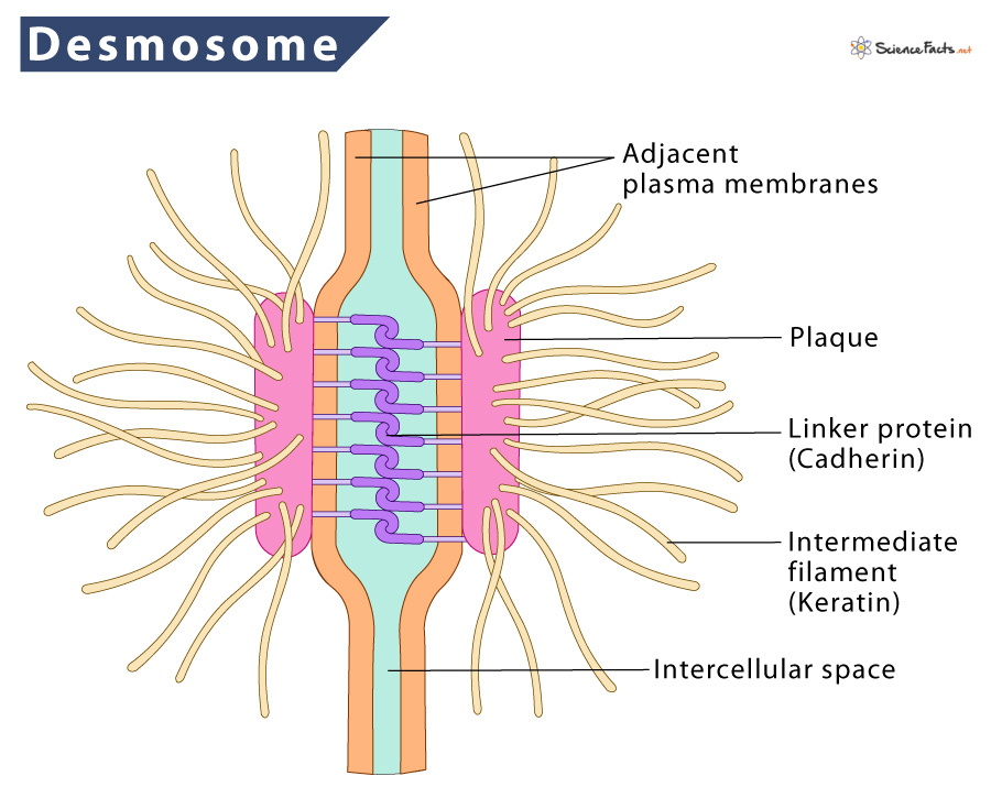
A DIFC consists of three parts:
1. Extracellular Core Region (Desmoglea) is approximately 34 nm in length containing the cadherin family of cell adhesion proteins – desmoglein and desmocollin.
2. Outer Dense Plaque (ODP) is about 15–20 nm in length containing the intracellular ends of desmocollin and desmoglein, the N-terminus portion of desmoplakin, and the armadillo family proteins plakoglobin and plakophilin.
3. Inner Dense Plaque (IDP) is about 15–20 nm in length containing the C-terminus portion of desmoplakin and their attachment to keratin intermediate filaments. Desmoplakin is most abundant in desmosome, acting as the moderator between the cadherin proteins in the plasma membrane and the keratin filaments
Functions of Desmosomes
The primary function of desmosomes is to provide strong adhesion between adjacent cells, acting like spot welds holding the tissues together. They mediate direct cell-cell contacts and provide anchorage sites for intermediate filaments essential for maintaining tissue integrity.
Since desmosomes connect intermediate filaments of the cell cytoskeletons of adjacent cells, they provide mechanical strength to tissues. Also, they are found to affect the cadherin protein-based intracellular signaling pathways.
Clinical Significances of Desmosomes
Malfunctioning of desmosomes is often the cause of certain human diseases, such as arrhythmogenic cardiomyopathy and blistering diseases.
Arrhythmogenic cardiomyopathy is usually caused due to mutations in desmoglein 2, but sometimes in desmocollin 2. Blistering disease such as pemphigus vulgaris (PV) is an autoimmune disease where auto-antibodies target desmogleins. PV is caused by circulating autoantibodies (IgG) that target Dsg3 (Desmoglein 3) and sometimes Dsg1.
FAQs
Ans. Cardiac muscle cells have desmosomes and gap junctions.
-
References
Article was last reviewed on Friday, August 19, 2022
Related articles
Popular Articles


Join our Newsletter
Fill your E-mail Address