Fluid Mosaic Model
The fluid mosaic model is a way biologists use to describe the structure of biological membranes, such as the cell membrane. It was first proposed by Seymour Jonathan Singer and Garth L. Nicolson in 1972. The model has been modified in parts over time, keeping the basic concept the same.
It describes the structure of the cell (plasma) membrane as a mosaic of components – proteins, phospholipids, carbohydrates, and cholesterol giving the membrane a fluid character. However, the proportion of the components varies with cell type. For example, the inner mitochondrial membrane contains 76% protein and 24% lipid, while myelin has 18% protein and 76% lipid in its membrane.
Fluid Mosaic Model and Cell Membrane
According to the fluid mosaic model, the main components of the cell membrane are:
- Phospholipids: The fabric of the membrane forms a bilayer with phosphate heads facing outwards and fatty acid tails facing inwards. They are amphiphilic or dual-loving.
- Cholesterol: A steroid that regulates the entry and exit of molecules in and out of the cell. It is attached to a single phospholipid and between the two phospholipid layers.
- Proteins: Based on the position in the membrane, they are of two types: integral and peripheral proteins.
- Carbohydrates: They are attached to proteins on the outside.
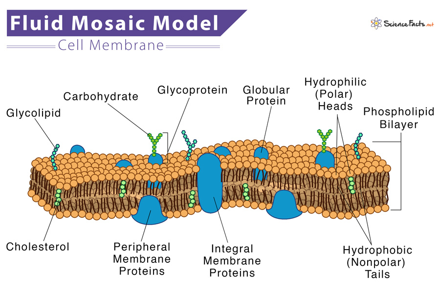
The model describes the structure of the cell membrane as a mosaic of these components having a thickness of 5-10 nm.
Phospholipids
Phospholipids are amphiphilic, having water-attraction (hydrophilic) and water-repulsion (hydrophobic) regions. The polar, hydrophilic head is constantly in contact with the fluid inside and outside the cell. In contrast, the non-polar, hydrophobic tail is away from the aqueous environment. Each phospholipid molecule has a glycerol backbone with two fatty-acid molecules and a phosphate group attached to it.
In an aqueous environment, the hydrophobic molecules arrange themselves to form a ball or a cluster. Hydrophilic regions form hydrogen bonds with water and other polar molecules. Thus the surface of the cell membrane facing the interior and exterior of the cell is hydrophilic. This arrangement allows phospholipid molecules to form a bilayer structure that separates the cell’s interior from the exterior.
Cholesterol
Cholesterol is found within the phospholipids, allowing the membrane to retain its permeability and integrity even if there is a temperature change. Cholesterol also prevents the compaction of the hydrophobic tails of lipids at low temperatures and the membrane’s expansion under heat. It helps the cell membrane selectively permeable to larger molecules while allowing small molecules like carbon dioxide and oxygen to pass through easily.
Proteins
Proteins are the second major component of the cell membrane and are randomly arranged within the lipid bilayer. Integral proteins are entirely integrated into the membrane structure. Their hydrophobic membrane-spanning part interacts with the hydrophobic part of the bilayer.
Single-pass integral proteins have a single membrane-spanning region consisting of 20–25 amino acids. Some integral proteins span only part of the membrane. In contrast, others have a transmembrane region from one side to the other. In contrast, multi-pass proteins transverse the membrane multiple times that are folded and embedded in the membrane. Thus, this type of membrane protein has a hydrophilic region(s) and one or several mildly hydrophobic regions.
This arrangement of proteins tends to orient them along with the phospholipids, with the protein’s hydrophobic region adjacent to the phospholipid’s tails and the hydrophilic regions protruding from the membrane, having contact with the cytosol or the extracellular fluid.
Carbohydrates
Apart from the three main parts, a cell membrane contains carbohydrates on the exterior surface of cells attached to proteins (forming glycoproteins) or lipids (forming glycolipids). Each carbohydrate chain consists of 2-60 monosaccharide units. With peripheral proteins, carbohydrate chains form specialized sites that allow cell-cell recognition. These carbohydrates on the exterior cell surface in the glycoproteins and glycolipids are together called the glycocalyx. It is hydrophilic and attracts water to the cell surface and thus helps to absorb substances dissolved in water.
All these components are arranged to give it a mosaic pattern. Scanning electron microscope images show that the embedded molecules, such as proteins and lipids, can move sideways through the membrane, which means the membrane is fluid-like and not solid. Such movement causes a change in the mosaic pattern of the cell membrane. With the help of proteins, lipids, and carbohydrates, the cell constantly interacts with the outside environment, thus allowing the import of ions, hormones, and food inside the cell and exporting waste products.
What does Fluid Mosaic Model Do
The fluid mosaic model also helps to recognize the functions of the different components in the cell membrane. It also explains how the membrane functions as a barrier between the cell cytosol and the extracellular fluid and develop cell-cell communication.
-
References
Article was last reviewed on Wednesday, February 1, 2023
Related articles
Popular Articles


Join our Newsletter
Fill your E-mail Address