Gap Junctions
Gap junctions are a group of intercellular channels that establish direct connections between neighboring cells in animals. They are also known as communicating junctions, macula communicans, or nexuses. They are one of the four categories of cell-cell junctions, along with tight junctions, adhering junctions, and desmosomes.
Similar to plasmodesmata in plant cells, gap junctions allow the passing of ions, small molecules, and electrical signals between two or more animal cells, facilitating communication between them.
Where are Gap Junctions Found
They are located in various animal tissues and organs, such as the nervous system, cardiac muscle, smooth muscle, epithelial tissues, connective tissue, and reproductive system.
- In the nervous system, gap junctions are found in regions like the brain, spinal cord, and peripheral nerves.
- The intercalated discs, specialized regions between adjacent cardiomyocytes in cardiac muscle, contain numerous gap junctions.
- In smooth muscle, they line the walls of various organs, such as the digestive tract, blood vessels, and reproductive organs.
- The epithelial cells covering the external body surface and lining the internal organs contain gap junctions.
- Certain types of connective tissues, such as bone and cartilage, also contain gap junctions.
- Even gap junctions exist in reproductive tissues, including the ovaries, testes, and placenta.
Structure of Gap Junctions
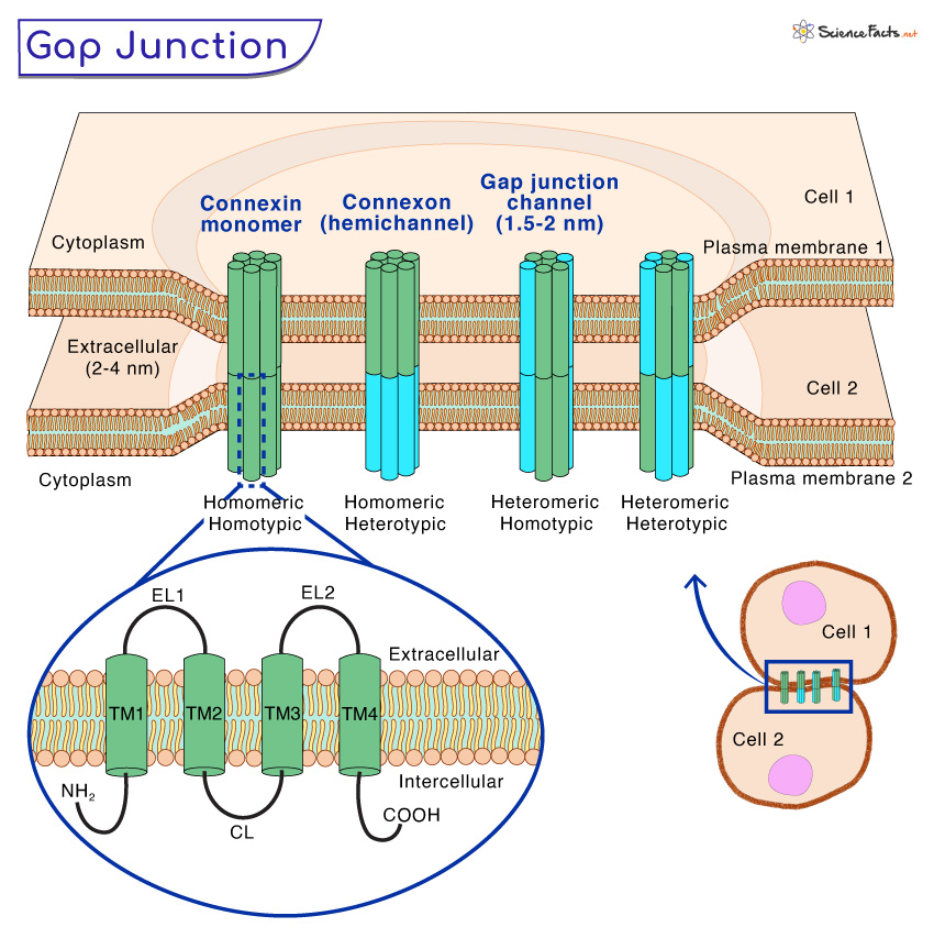
Gap junctions consist of two hemichannels, also known as connexons, embedded in the plasma membranes of adjacent cells with a 2-4 nm gap between them. Each connexon is composed of six connexin protein subunits forming a hexamer. Connexins are a family of transmembrane proteins involved explicitly in forming gap junctions.
The connexin subunits form a cylindrical structure with a central pore. Each subunit consists of four transmembrane domains (TM1 to TM4), two extracellular loops (EL1 and EL2), a cytoplasmic loop (CL), and a cytoplasmic amino-terminal (NT) and a carboxyl-terminal (CT).
The connexin subunits within a connexon align perfectly, creating a channel that spans the intercellular space between the two neighboring cells. The channel formed by the connexons is known as a gap junction channel or gap junction pore. The connexin subunits exhibit diversity and combinations that give rise to various connexon types with distinct properties.
The central pore of the gap junction channel is approximately 1.5-2 nm in diameter. This pore allows the passage of small molecules between the connected cells, including ions, metabolites, and signaling molecules.
Connexons consisting of a single connexin type are called homomeric, and those composed of more than one connexin type are called heteromeric connexons. When connexons of the same composition form a gap junction channel, it is termed homotypic. If the connexons differ in components, the channel is termed heterotypic.
Assembly of Gap Junctions
The assembly of gap junctions is regulated by various mechanisms that are achieved through the following steps:
1. Synthesis of Connexin Proteins
The first step in gap junction assembly begins with synthesizing connexin proteins in the cell’s endoplasmic reticulum (ER). Connexins are a family of transmembrane proteins comprising the gap junction channels. Each connexin protein comprises four transmembrane domains, two extracellular loops, a cytoplasmic loop, and cytoplasmic amino and carboxyl termini.
2. Oligomerization of Connexins
Once synthesized, individual connexin proteins undergo a process known as oligomerization. Six connexin subunits form a hexameric structure called a connexon or hemichannel. Each connexon represents half of the future gap junction channel.
3. Post-translational Modifications
As the connexons are assembled, they undergo post-translational modifications, such as phosphorylation, glycosylation, and palmitoylation. These modifications are crucial for regulating the stability, trafficking, and functional properties of the connexons.
4. Trafficking to the Plasma Membrane
After oligomerization and post-translational modifications, the connexons are moved from the endoplasmic reticulum to the Golgi apparatus and the cell’s plasma membrane. This trafficking process is tightly regulated and ensures that the connexons reach their appropriate destinations.
5. Docking of Connexons
At the plasma membrane, the connexons from one cell align with connexons from a neighboring cell. These opposing connexons come into proximity, and with the help of specific proteins, they dock together, forming the complete gap junction channel. This docking is a critical step in the assembly process, as it creates a continuous channel connecting the cytoplasm of adjacent cells.
6. Formation of Gap Junction Channel
Once the connexons are docked, the gap junction channel is formed. The channel spans the intercellular space, allowing direct communication between the cytoplasm of the connected cells. The central pore of the gap junction channel, approximately 1.5 to 2 nanometers in diameter, permits the passage of ions, metabolites, and small signaling molecules.
Functions of Gap Junctions
Gap junctions perform numerous functions in cells. They are discussed below:
Direct Cell-to-Cell Communication
Gap junctions allow direct communication between neighboring cells. It allows them to exchange various substances, including ions, metabolites, and signaling molecules. This direct transfer of substances facilitates coordinated and synchronized cellular activities within tissues.
The size of the molecules that pass through the channel is restricted by the pore’s diameter and the specific connexin types in the gap junction.
Electrical Synchronization
Gap junctions are crucial for the electrical synchronization of cells in tissues that exhibit electrical activity, such as cardiac muscle and neurons. In the heart, for example, gap junctions allow the rapid propagation of electrical signals, known as action potentials, from one cardiac cell to the next. This synchronized electrical activity ensures coordinated contraction and proper functioning of the heart.
Metabolic Cooperation
Gap junctions facilitate metabolic cooperation between cells. They enable the exchange of metabolites, such as glucose, amino acids, and nucleotides, ensuring the efficient distribution of nutrients and removing waste products. This metabolic cooperation is essential in tissues with high energy demands, such as the liver and brain.
Tissue Development and Differentiation
Gap junctions are crucial in cell signaling and tissue organization during embryonic development. They contribute to cell differentiation, cell migration, and the establishment of tissue-specific functions. Gap junction-mediated communication helps coordinate the activities of different cell types, ensuring proper development and organization of tissues and organs.
Homeostasis and Tissue Function
Gap junctions are essential for maintaining tissue homeostasis. By allowing neighboring cells to communicate and synchronize their activities, these intercellular channels help regulate tissue functions.
Tissue Repair and Regeneration
During tissue repair processes, such as wound healing, gap junctions play a role in coordinating cellular responses. They facilitate the transfer of signaling molecules, allowing cells to communicate and coordinate their actions to promote tissue regeneration.
Cell Signaling and Coordination
Gap junctions participate in intercellular signaling and coordination. They facilitate the exchange of various signaling molecules, including secondary messengers and small regulators. This intercellular signaling contributes to coordinating cellular responses and allows cells to communicate important information to neighboring cells.
Regulation of Gap Junction Functions
The function of gap junction channels is highly regulated to adapt to the specific needs of the cells and tissues. Various factors, such as changes in calcium concentration, pH levels, or the phosphorylation state of connexins, can modulate the opening and closing of gap junction channels. This regulation allows for precise control of intercellular communication and coordination of cellular activities.
Turnover and Degradation of Gap Junctions
Gap junction channels are dynamic, with connexins continuously being synthesized, assembled, and degraded. The turnover of gap junction proteins ensures the maintenance of functional channels. It allows cells to adapt their communication needs in response to physiological changes.
Gap Junctions and Disorders
Defects in gap junctions can lead to various diseases and disorders. For example, mutations in connexin genes can result in conditions like Charcot-Marie-Tooth disease, which affects peripheral nerves, or oculodentodigital dysplasia, which affects multiple organs. Disrupted gap junction communication has also been implicated in cardiovascular diseases, cancer, and neurological disorders.
FAQs
Ans. Intercalated discs in cardiac muscle contain both gap junctions and desmosomes.
Ans. Skeletal muscles do not have gap junctions.
-
References
Article was last reviewed on Tuesday, August 29, 2023
Related articles
Popular Articles


Join our Newsletter
Fill your E-mail Address