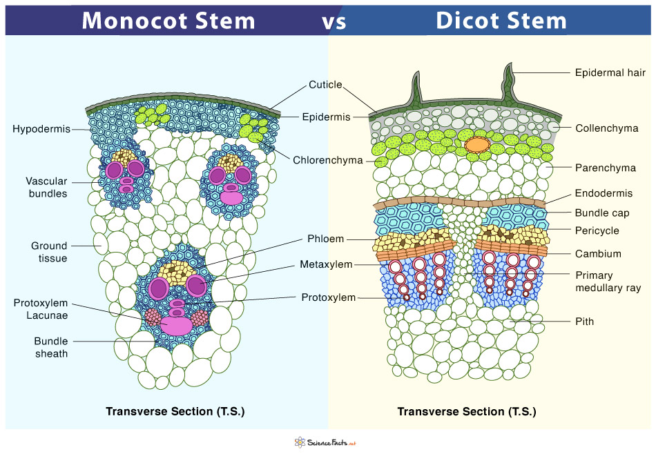
Monocot vs Dicot Stem
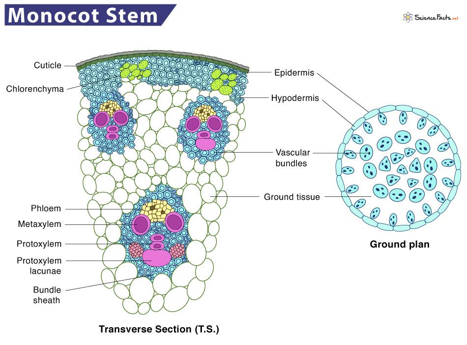
The monocot stems are stems of monocot plants. They have scattered vascular bundles of xylem and phloem, surrounded by a bundle sheath of sclerenchyma cells. The monocot stems have other significant features: lack of trichomes (epidermal hairs), medullary rays, cortex or pith, and a stele. Also, the hypodermis consists of sclerenchyma cells.
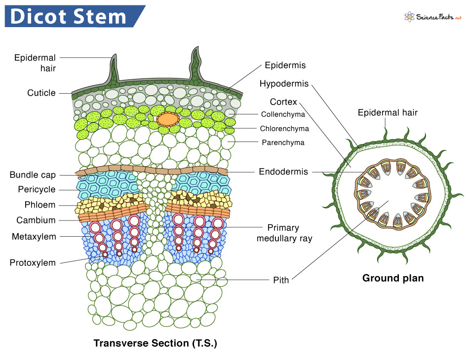
The dicot stems are stems of dicot plants. They are arranged concentrically, one above the other. The vascular bundles of dicot stems are arranged in a ring without a bundle sheath. However, parenchyma cells surround each vascular bundle in the dicot stem. The hypodermis of dicot stems is made of collenchymas cells. They have other significant features such as trichomes in the epidermis, prominent cortex, and stele. In addition, dicot stems show secondary thickening, which leads to secondary growth.
Let us discuss the structural and functional similarities in monocot and dicot plants.
Similarities
Structure
The internal structure or anatomy of the stems of monocot and dicot plants has the following parts in common.
Epidermis
- It is the single, outermost layer arranged compactly without intercellular spaces.
- The epidermis cells lack chlorophyll
- Outside the epidermis is a layer of the cuticle that protects the epidermis and tissues lying underneath
- It contains stomata for the gas exchange
Cortex
- It is divided into different sections, but the region covered differs widely between monocot and dicots stems.
- The hypodermis is present immediately below the epidermis, which provides additional support to the epidermis. It is a thick multicellular layer composed of collenchyma tissue. The cells of this region contain chlorophyll and thus can prepare food for the plant.
Ground Tissue
- It is a mass of parenchymatous cells extending to the hypodermis center.
- The cells are spherical, thin-walled, and loosely organized with intercellular spaces.
Pericycle
- It is the tissue found between the endodermis and the vascular bundles.
- The cells are sclerenchymatous, which has the shape of semilunar patches above the vascular bundles.
Vascular Bundles
- They are of conjoint and collateral type surrounded by parenchyma cells
- They are limited in number and have a uniform size. Each bundle consists of a patch of xylem and phloem
- Xylem consists of both protoxylem and metaxylem
- The quantity, arrangement, and components of vascular bundles vary widely between monocot and dicots stems
Pith
- The cells are parenchymatous and appear like medullary rays around the arterial bundles.
- The cells might be spherical or polygonal, with or without any intercellular spaces.
- They store food and aid in transporting food and water between the bundles.
Functions
Both monocot and dicot stems perform the same essential functions that are discussed below:
- It is the central axis of the plant that supports other parts such as leaves, branches, flowers, and fruits
- Transports food, water, and minerals throughout the plant body
- Young, green stem having pigments chlorophyll prepares food by photosynthesis
- Stores a large number of food particles like starch and other nutrients
- The meristem tissue of the stem divides to form new tissues, thus helping the plant to grow
- The stomata in the stem helps in transpiration
- Modified stems like cactus help to store water and food, prevent water loss, and thus in their survival in the desert
Although monocot and dicot stems have the following structures in common, there are several differences. They are discussed below.
Difference between Monocot and Dicot Stem
| Basis | Monocot Stem | Dicot Stem |
|---|---|---|
| Examples | Tulips, onions, lilies, and garlic | Pea, cactus, sunflower, and walnut |
| Epidermis | It remains the same throughout the growth of the plant | Replaces during secondary growth |
| Epidermal Hairs | Present over the epidermis | Absent |
| Vascular Bundles | 1. Conjoint, collateral, and closed type 2. Remain unchanged throughout the life of the plant 3. Numerous 4. Scattered across the stem 5. Outer vascular bundles are smaller than the inner ones 6. Do not contain a sclerenchymatous bundle cap 7. Surrounded by a sclerenchymatous bundle sheath 8. Xylem elements are circular 9. The xylem contains protoxylem lacunae 10. Has only two metaxylem elements per vascular bundle 11. Phloem units are smaller in size than dicots 12. Phloem parenchyma and phloem fibers absent | 1. Conjoint, collateral, and open type 2. Old vascular bundles are replaced by new ones many times throughout the plant life 3. 4 to 8 4. Arranged in the form of rings 5. All vascular bundles are of equal size 6. Contain a sclerenchymatous bundle cap 7. Not surrounded by a sclerenchymatous bundle sheath 8. Xylem elements are polygonal 9. Does not have protoxylem lacunae 10. Has many metaxylem elements per vascular bundle 11. Phloem units are larger than monocots 12. Phloem parenchyma and phloem fibers are present |
| Pith | Not as well-developed as dicots | Well developed |
| Ground Tissue | It is undivided into units such as pericycle and pith | It is divided into pericycle, medullary rays, and pith |
| Cortex | 1. Less developed and is differentiated only into the hypodermis 2. The hypodermis is sclerenchymatous and mostly non-green 3. The hypodermis is stiffer than dicots 4. The general cortex is reduced or absent 5. The endodermis is absent | 1. Well-developed and differentiated into hypodermis, endodermis, and general cortex 2. The hypodermis is collenchymatous and mostly green 3. The hypodermis is more flexible than monocots 4. The general cortex is present 5. The endodermis is present |
| Stele | 1. Larger than dicots 2. Advanced type 3. It consists of ground tissues and vascular bundles | 1. Smaller than monocots but larger than the cortex 2. Moderately developed 3. Differentiated into pericycle, vascular medulla, and medurally rays |
| Pericycle | Reduced or completely absent | Present, partially, or completely sclerenchymatous |
| Trichomes | Absent | Present |
| Secondary Growth | Not found | Found due to the presence of secondary vascular tissues and periderm formation |
| Medullary Rays | Absent | Present |
| Internodes | Hollow | Solid |
-
References
Article was last reviewed on Thursday, February 2, 2023
Related articles
Popular Articles


Join our Newsletter
Fill your E-mail Address