Microfilaments
What are Microfilaments
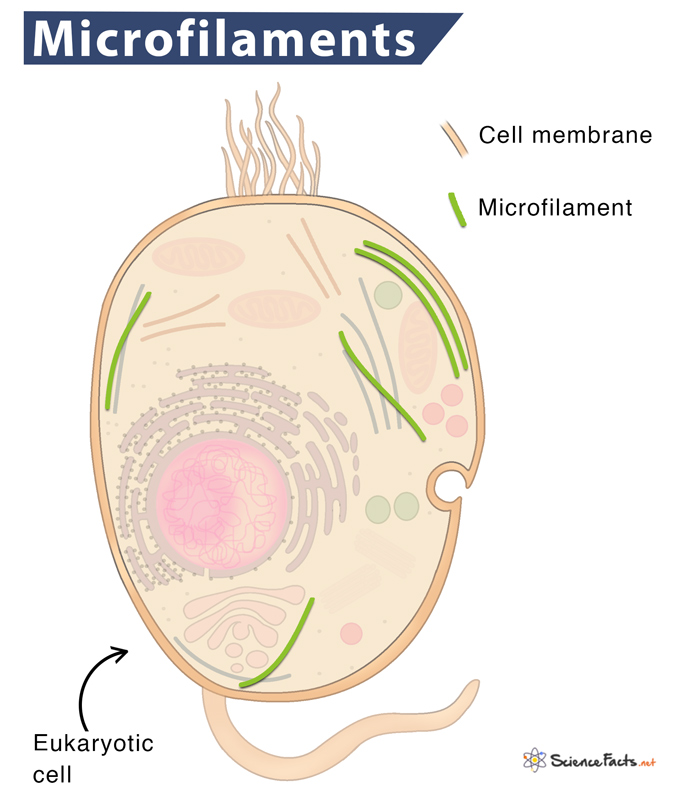
Microfilaments, also known as actin filaments, are actin proteins arranged in a long, spiral chain. They are the narrowest of the three cytoskeletons found in the cytoplasm of all eukaryotic cells. Microfilaments help to maintain cell shape.
Location
They are typically nucleated beneath the plasma membrane and are part of the cell cortex. Thus, the periphery of a cell generally contains the highest concentration of microfilaments, where they attach to the plasma membrane and reach the microvilli.
The red blood cells (RBCs), human embryonic kidney cells, neurons, and sperm cells are examples of specialized unique actin cytoskeletal structures adjacent to the plasma membrane.
Structure
Microfilaments are long, thin, and stringy proteins. When first produced by the cell, the actin monomers join together to form two parallel polymers of globular-(G)-actin. Once they are joined, the elongated strands twist around each other into a helical orientation having a diameter of about 6-7 nm and are called filamentous-(F)-actin.
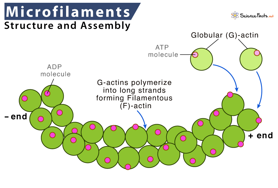
Like microtubules, all the subunits of microfilaments are connected in the same orientation, thus exhibiting polarity having a positive or plus (+) end and a negative or minus (-) end. The positive or the fast-growing end is barbed, while the negative or slow-growing end is pointed.
The actin filaments within the cells are usually organized into more extensive, robust structures with accessory proteins. The structural form that a group of microfilaments develops depends on their primary function and the type of proteins they associate.
Formation
The self-assembly of microtubules begins to form when three G-proteins come together to form a trimer. Subsequently, more actin binds to the plus end in a step-by-step manner forming long strands of globular actin. The two long strands wound spirally around each other to form a microfilament. The entire process is ATP-dependent.
Microfilaments can also depolymerize (disassemble) and reform quickly, thus enabling a cell to change its shape and move. The continuous removal of actin monomers from the pointed ends of filaments and their reincorporation at barbed ends is called actin treadmilling.
Functions
1. Cell Division
Together with myosin, microfilaments play a crucial role in cell division. During cytokinesis, a ring of actin forms around the cell that separates. Myosin pulls on the actin and causes it to contract. The ring gets narrower around the cell dragging the cell membrane with it until the two cells ‘pinches off’ and separate into two daughter cells.
2. Muscle Contraction
The protein actin works together with myosin to generate the forces required for the contraction and relaxation of muscles. Thus, the amount of microfilaments in muscle cells is high. They form myofibrils, the basic unit of the muscle cell.
3. Cell Movement
They play a crucial role in causing eukaryotic organisms to move from one place to another. They are also crucial in the movement of single-celled prokaryotes such as amoeba and paramecium (amoeboid movement). For movement, one end of the microfilament elongates while the other end shortens. This assembly and disassembly occur with the help of myosin.
4. Cytoplasmic Streaming
They allow the flow of cytoplasm (the cell content, including cytosol and organelles) within the cell. Microfilaments thus help nutrients, waste products, and cell organelles travel from one part of the cell to the other part. Also, they attach to cell organelles and contract, pulling the organelles to a different area.
5. Forming Cell Surface Projections
Microfilaments play a vital role in developing various cell-surface projections such as filopodia, lamellipodia, and stereocilia. These structures act as antennae for a cell to probe the surrounding environment.
6. Cell Stability and Survival
All eukaryotic cells depend on the actin filaments’ integrity to survive the stress of the outside environment. Accordingly, many plants that cannot avoid predators mechanically produce toxins that affect actin and microfilaments as a defensive mechanism.
7. Fertilization
They are involved in sperm incorporation in animals. Microfilaments also help in spindle rotation (in mice), cortical granule exocytosis, second polar body emission, and cleavage ring formation during fertilization.
-
References
Article was last reviewed on Tuesday, February 8, 2022
Related articles
Popular Articles


Join our Newsletter
Fill your E-mail Address