Microtubules
Microtubules are one of the three principal components of the cytoskeleton, along with microfilaments and intermediate filaments. They are found throughout the cytoplasm in eukaryotic plant and animal cells. Like the other two cytoskeletal elements, microtubules play a crucial role in cell division, movement, and maintaining cell shape.
In most cells, microtubules extend outward from the microtubule-organizing centers (MTOCs). In animal cells, the major microtubule-organizing center is the centrosome, located adjacent to the nucleus. Microtubules are also located in cilia and flagella of prokaryotes and human sperm cells.
Structure of Microtubules
Size
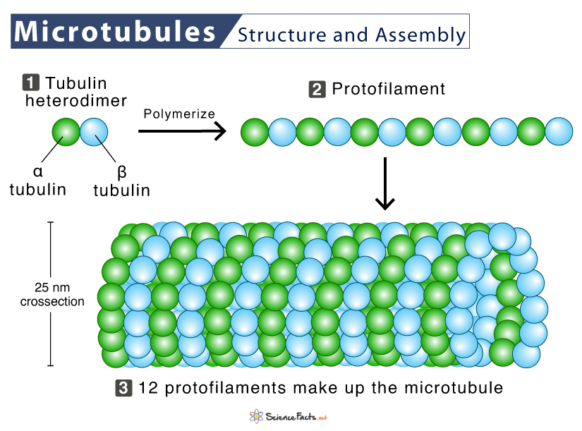
They are rigid hollow rods of approximately 25 nm in diameter and between 200 nm and 25 μm in length. Microtubules consist of a single type of cytoplasmic, globular protein subunits called tubulin.
Composition
Each microtubule subunit comprises two closely related polypeptides: α-tubulin, and β-tubulin, forming heterodimers. Multiple units of these dimers polymerize to form a chain called the protofilament. Then, 13 protofilaments arrange into a cylindrical pattern to form a microtubule.
In a microtubule, the subunits are organized such that they all face the same direction to form 13 parallel protofilaments. Thus, the microtubule is polar with the alpha-tubulin exposed at one end and beta-tubulin at the other.
Assembly and Disassembly of Microtubules
- Since microtubules are polar, they have a positively charged or (+) end that grows relatively fast and a negatively charged or (-) end that grows relatively slowly.
- Each protofilament arranges in a parallel orientation concerning the other such that the positive end always has beta subunits exposed. In contrast, the negative end has alpha subunits exposed.
- Tubulin dimers can also depolymerize. Both α- and β-tubulin bind GTP to power polymerization or depolymerization. In particular, the GTP bound to β-tubulin is hydrolyzed to GDP during polymerization. Hydrolysis weakens the binding affinity of tubulin for adjacent molecules, favoring depolymerization and resulting in the dynamic behavior of microtubules.
Microtubules and Treadmilling
Like actin filaments, microtubules undergo treadmilling. Here, the tubulin molecules bound to GDP are continuously lost from the minus end and replaced by their addition bound to GTP to the plus end of the same microtubule.
Microtubules and Dynamic Instability
When new GTP-bound tubulin molecules add up more rapidly than GTP is hydrolyzed, the microtubule holds on to a GTP cap at its plus end, causing the microtubule to grow. In contrast, if the rate of polymerization decreases, then the GTP bound to tubulin at the plus end is hydrolyzed to GDP. Finally, the GDP-bound tubulin dissociates, resulting in rapid depolymerization and shrinkage of the microtubule. This phenomenon where microtubules alternate between cycles of growth and shrinkage is called dynamic instability.
Functions of Microtubules
Some of the main functions of microtubules are:
Maintaining Cell Shape
Microtubules help maintain cell shape and stability with microfilaments and intermediate filaments. Together with the other cytoskeleton element, microtubules form an architectural framework that establishes the overall polarity of the cell.
Cell Movement
Cells provide structure to cilia and flagella and thus help move bacteria and other prokaryotes. Motile cilia and eukaryotic flagella have the canonical ‘9+2’ arrangement of microtubules where nine doublet microtubules surround a central pair of singlet microtubules.
Microtubules in the trachea cells prevent mucus and dirt from entering the lungs. The fallopian tubes (female reproductive system) move the egg released from the ovary to the uterus.
Cell Division
During mitosis, microtubules play a crucial role in forming the mitotic spindle (spindle apparatus). The mitotic spindle helps to separate chromosomes during cell division so that the chromosomes can be partitioned equally into two daughter cells. The spindle apparatus also helps to form the contractile ring that separates the two daughter cells during cytokinesis.
Three types of microtubules participate in mitosis: astral, polar, and kinetochore microtubules. Astral microtubules radiate from the MTOCs of a cell to the cell membrane, thus keeping the mitotic spindle in place. Polar microtubules link between two MTOCs and help separate chromosomes. Kinetochore microtubules attach to chromosomes that help to pull them apart.
Intracellular Transport and Communication
As part of the cytoskeletal network, microtubules help move organelles inside a cell’s cytoplasm. Microtubules also help the various cell components to communicate with each other.
Forming an Internal Transport Network
They form an internal transport network for moving materials throughout the cell and between the exterior and interior of the cell. This trafficking is done by microtubule-associated proteins kinesin and dynein.
FAQs
Ans. The kinetochore microtubules are found at the centromere of chromosomes serving as a bridge between the chromosome DNA and the non-kinetochore microtubules. In contrast, non-kinetochore microtubules are the mitotic spindle fibers that help divide the chromosomes.
Ans. Microtubules are the smallest of the entire cytoskeletal element, with a diameter of 5 nanometers.
-
References
Article was last reviewed on Friday, February 17, 2023
Related articles
Popular Articles


Join our Newsletter
Fill your E-mail Address