Sarcoplasmic Reticulum
The Sarcoplasmic reticulum (SR) is a unique form of smooth endoplasmic reticulum of skeletal muscle cells. Like the endoplasmic reticulum of other cells, it is a network of closed sac-like membranes found distributed throughout the cell. It spans the sarcomere and encloses the contractile myofilaments in striated muscle cells.
The primary function of SR is to store and release calcium ions (Ca2+) during muscle contraction and relaxation.
Structure of Sarcoplasmic Reticulum
Studies with an electron microscope revealed that in skeletal muscles, the SR is organized into numerous interconnected tubules and sacs forming a network of longitudinal elements, known as longitudinal SR (l-SR), surrounding each myofibril. The SR is enclosed by sarcolemma, the cell membrane of muscle cells.
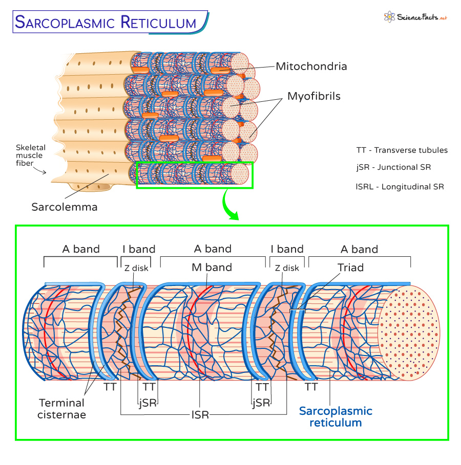
L-SR is found around each sarcomere’s anisotropic (A) and isotropic (I) bands. A and I bands are bisected by the M band and the Z disk, respectively, and the portion of the myofibril between two Z disks is the sarcomere. In the border between the A and I bands, the I-SR tubules merge into enlarged sac-shaped structures called terminal cisternae. The two terminal cisternae, called transverse tubules (T tubule or TT), are located at the opposite sides of the tubular infoldings, continuous with the nuclear envelope. The structure formed by two terminal cisternae and one TT is called a triad.
The SR has two distinct regions: the junctional SR (jSR) and the free SR (fSR), each with unique features. The jSR has an elongated, flattened appearance, with an average diameter measuring about 0.6 μm. Within the jSR, there are closely grouped structures called feet. These structures house the SR calcium release channels, called ryanodine receptors (RyRs). Additionally, the jSR contains a dense material composed of the calcium-binding protein calsequestrin-2 (CASQ2). In contrast, the fSR lacks CASQ2 and has an external surface adorned with densely distributed particles corresponding to calcium adenosine triphosphatase (ATPase).
Functions of Sarcoplasmic Reticulum
The SR is a membrane-bound structure within skeletal muscle cells that takes up, stores, and releases Ca2+. Calcium enters SR through specialized Ca2+ channels, an ATP-dependent process, while it exits ST through RyRs.
Role in Calcium Absorption
The calcium ion concentration inside the SR is higher than the rest of the cell. They require a pump similar to Na+/k+ ATPase, which pumps Na+ and k+ ions against their concentration gradient for more calcium absorption.
This active calcium absorption is done by a specialized calcium pump called the Sarco(endo)plasmic reticulum Ca2+ ATPases (SERCA).
Role in Calcium Storage
After calcium absorption, they need to be stored for the proper functioning of the cell. The storage is accomplished using a protein called calsequestrin, located at the junctional SR/luminal space in association with the calcium release channel. A single calsequestrin protein molecule can bind around 50 Ca2+, thus decreasing the amount of free Ca2+ within the SR.
Role in Calcium Release
Apart from calcium absorption and storage, the SR is also responsible for releasing calcium ions from the cell. It occurs in the junctional SR/terminal cisternae through the RyR receptor, a process called calcium spark.
Helps in Muscle Contraction
Calcium ions’ absorption, storage, and release help skeletal muscles contract and expand according to our muscular activities and needs. Calcium binds to proteins in the myofibrils, specifically troponin and tropomyosin, which regulate the contraction process. When calcium binds to these proteins, it allows the myosin and actin filaments to interact, resulting in the contraction of muscles.
Role of Sarcoplasmic Reticulum in Rigor Mortis
SR is responsible for releasing calcium into the cell cytoplasm, which is required for muscle contraction. Calcium, however, returns to SR after contraction and is stored. However, this step is ATP-dependent, which is obtained from aerobic respiration.
After death, the heart stops pumping oxygen, and thus, our body cannot produce ATP. In such a situation, the return of calcium ions into the SR prevents the accumulation of calcium ions within the cell, leading to prolonged contraction of muscles, a phenomenon known as rigor mortis.
Role of Sarcoplasmic Reticulum in Heart Diseases
As SR is mainly responsible for storing and releasing calcium in muscle cells, any defects in the calcium pump on SR will cause the Ca2+ ions to vary within the cell. A faulty SR is found to be a cause of heart failure. However, more research is needed to understand the role of ST in heart complications.
Sarcoplasmic Reticulum vs. Endoplasmic Reticulum
SR is analogous to the smooth endoplasmic reticulum (SER) of skeletal muscle cells and is responsible for their calcium uptake, storage, and release. On the other hand, the endoplasmic reticulum is responsible for protein synthesis, modification, secretion, lipid and steroid synthesis, and modulation of Ca2+ signaling.
-
References
Article was last reviewed on Monday, October 9, 2023
Related articles
Popular Articles


Join our Newsletter
Fill your E-mail Address