Hemidesmosomes
Hemidesmosomes (HD) are specialized anchoring junctions found in basal epithelial cells connecting them to their underlying basement membrane. They are crucial in tissues under constant stress, such as the skin, cornea, parts of the gastrointestinal and respiratory tracts, esophagus, amnion, and vagina. They are responsible for providing them with strength and rigidity. Together with intermediate filaments, hemidesmosomes provide the overall strength and stability of epithelial tissues.
They are also called half-desmosomes due to their structural resemblance to desmosomes, which are intercellular junctions that hold adjacent cells together. They are also compared to focal adhesion, as they attach cells to the extracellular matrix. However, hemidesmosomes utilize integrins instead of desmogleins and desmocollins in focal adhesions.
Structure of Hemidesmosomes
Hemidesmosomes are made of several protein components. Ultrastructurally, they were characterized as unique structures with a dense appearance when viewed under an electron microscope. They consist of an internal plaque that connects to intermediate filaments within the cell, an external plaque that attaches to the extracellular basement membrane, and a dense plate beneath the outer plaque in the lamina lucida of the basement membrane. Hemidesmosomes can be either type 1 or type 2, based on their protein constituents.
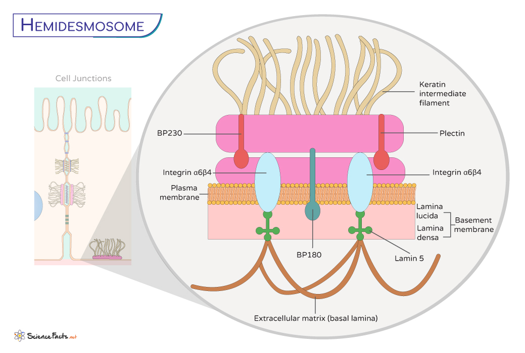
The main components of hemidesmosomes are summarized below with their positions in the cell-cell network:
- The transmembrane linker protein, integrin, is at its core, which spans the cell membrane and connects the extracellular matrix (ECM) components to the intracellular cytoskeleton. Integrin α6β4 forms bridges between the cell and the lamina densa of the basement membrane, creating a stable connection. On the intracellular side, the cytoplasmic tail of integrins binds to various cytoskeletal components, such as the keratin intermediate filaments, reinforcing the structure and providing stability.
- Collagen XVII, known as bullous pemphigoid antigen isoform – BPAG2 (BP 180), is also a transmembrane protein that contributes to the overall strength of the hemidesmosome attachment.
- In addition to integrins and collagen XVII, hemidesmosomes contain other crucial proteins such as BP230 and plectin. BP230 is essential for anchoring integrins to the intermediate filaments. At the same time, plectin serves as a linker between integrins and intermediate filaments.
Functions of Hemidesmosomes
Hemidesmosomes are like molecular anchors that maintain tissue integrity by establishing connections between epithelial cells and the underlying basement membrane.
Maintaining Tissue Integrity and Stability
The primary function of hemidesmosomes is to provide structural stability to tissues. They act as glue that firmly attaches epithelial cells to the underlying basement membrane, forming a protective outer layer of various organs. This connection is crucial in tissues subjected to abrasion and mechanical stresses, such as the skin and mucous membranes lining the oral cavity and the gastrointestinal tract.
In tissues like the skin, which are exposed to frequent stretching and movement, hemidesmosomes act as resilient bonds that resist the tendency of cells to pull away from the basement membrane. This anchoring is vital for maintaining the integrity of the epithelial barrier.
Providing Protection against Foreign Invaders
In the epidermis, hemidesmosomes work alongside tight junctions and desmosomes to establish a seamless barrier against external agents. This barrier prevents pathogens, toxins, and other harmful substances from infiltrating deeper tissues.
Participating in Cellular Signaling and Communication
The integrins within hemidesmosomes can transmit signals bidirectionally – outside-in (from the ECM to the cell) and inside-out (from the cell to the ECM). These bidirectional signaling pathways are critical in cellular processes like adhesion, migration, proliferation, and tissue repair.
Wound Healing and Tissue Regeneration
When tissues are damaged, as in wounds or injuries, hemidesmosomes are involved in wound healing and tissue regeneration. They help guide migrating epithelial cells to cover the wound, ensuring the newly formed tissue maintains proper attachment to the basement membrane.
Hemidesmosomes and Associated Diseases
Dysfunctional hemidesmosomes have been implicated in various diseases. Autoimmune conditions like pemphigoid involve the immune system attacking the components of hemidesmosomes, leading to blistering and detachment of the epithelial layer. Understanding the mechanism of their work is crucial for developing targeted therapies.
Desmosomes vs. Hemidesmosomes
Desmosomes and hemidesmosomes are anchoring junctions important for maintaining adherence within epithelial tissues. However, they are structurally and functionally different.
| Basis | Desmosomes | Hemidesmosomes |
|---|---|---|
| 1. What Is It | They are intercellular junctions connecting adjacent cells in epithelial tissues. | They are intercellular junctions connecting cells to the extracellular matrix (ECM). |
| 2. Structure and Parts | They consist of transmembrane glycoproteins called desmogleins and desmocollins and intracellular proteins like plakoglobin and desmoplakin. | They consist of transmembrane integrins, such as α6β4 integrin, and intracellular proteins, like BP180, BP230, and plectin. |
| 3. Location | Typically, they are found on the lateral sides of epithelial cells such as skin and heart. | They are generally prevalent in tissues like the skin, oral mucosa, and other epithelial linings. |
| 4. Main Function | They create strong adhesions between neighboring cells, helping tissues withstand stretching and contraction. | They prevent the detachment of cells to their extracellular matrix during mechanical stress. |
-
References
Article was last reviewed on Thursday, November 30, 2023
Related articles
Popular Articles


Join our Newsletter
Fill your E-mail Address