Anchoring Junctions
Anchoring junctions are one of the three main types of cell junctions widely distributed in animal tissues, along with tight and gap junctions. They are highly dynamic structures that attach cells to their neighbors and the extracellular matrix. Anchoring junctions are abundant in tissues subjected to severe mechanical stress, such as the heart, muscle, and epidermis.
Functions
Apart from anchoring, they also play critical roles in:
- Stabilizing the position of the cell, providing stability and rigidity, and supporting tissue integrity by holding cell sheets together
- Forming a tight seal around the neighboring cells restricting the flow of molecules between cells and from one side of the tissue to the other
- Regulating the mobility of the cells and also the tissues in their extracellular matrix through their substrates
Basic Structure
They have two main structural features:
- Transmembrane cell-adhesion molecules (CAMs) or adhesion receptors such as cadherins and integrins link the lateral surfaces of one cell to another or the basal surfaces of the cell to the extracellular matrix.
- Intracellular adaptor proteins such as vinculin, α-actinin, and filamin connect the adhesion molecules to the cytoskeleton.
In addition to transmembrane and intracellular adaptor proteins, some anchoring junctions also contain intracellular signaling proteins that enable the junctions to interact and send signals to the inside of the cell.
Types of Anchoring Junctions
Anchoring junctions occur in functionally two different forms:
- Adhering Junctions and Desmosomes hold cells together and are formed by transmembrane adhesion proteins belonging to the cadherin family.
- Focal Adhesions and Hemidesmosomes bind cells to the extracellular matrix. They are formed by transmembrane adhesion proteins of the integrin family.
Accordingly, there are three main types of anchoring junctions.
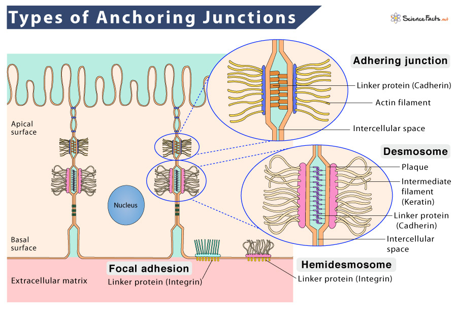
1. Adhering Junctions (Zonula Adherens)
They are cell-cell anchoring junctions that use actin filaments as their cytoskeletal anchor. Adhering junctions use cadherin or integrin as their transmembrane linker. While cadherin helps the cell anchor to other cells, integrin helps anchor them to the extracellular matrix.
- Adhering Junctions are found in epithelial and endothelial tissues.
2. Desmosomes (Macula Adherens)
They are another group of cell-cell anchoring junctions that use intermediate filaments like keratin as their cytoskeletal anchor. Desmosomes use cadherin as their transmembrane linker, providing strong adhesion between adjacent cells and thus acting like spot welds holding the tissues together.
- Desmosomes are found in tissues that experience stress, such as cardiac muscle tissue, bladder tissue, gastrointestinal mucosa, and epithelia.
3. Focal Adhesions and Hemidesmosomes
Unlike adhering junctions and desmosomes, focal adhesions bind cells to the extracellular matrix rather than other cells. The transmembrane adhesion proteins in these cell-matrix junctions are integrins. They enable cells to get a hold of the extracellular matrix through integrins that link intracellularly to actin filaments.
- Focal adhesions enable muscle cells to attach to tendons at the myotendinous junction.
Like focal adhesions, hemidesmosomes are cell-matrix junctions that use intermediate filaments similar to desmosomes as their cytoskeletal anchor. However, unlike them, hemidesmosomes connect the basal surface of an epithelial cell to the underlying basal lamina using integrin as the transmembrane linker.
- Hemidesmosomes are found only in epithelial cells.
-
References
Article was last reviewed on Friday, August 19, 2022
Related articles
Popular Articles


Join our Newsletter
Fill your E-mail Address