Meiosis
What is Meiosis
Meiosis is a cell division process where a single (parent) cell divides twice to produce four independent (daughter) cells, each having half the chromosomes as the original cell.
The term ‘meiosis’ came from the Greek word ‘meiosis’, meaning ‘lessening’.
Where does Meiosis Occur
Meiosis takes place only in the reproductive cell types (sperm and egg cells) of sexually reproducing organisms, including humans. For a cell to undergo meiosis, it must have a diploid (2n) chromosome number.
Phases of Meiosis
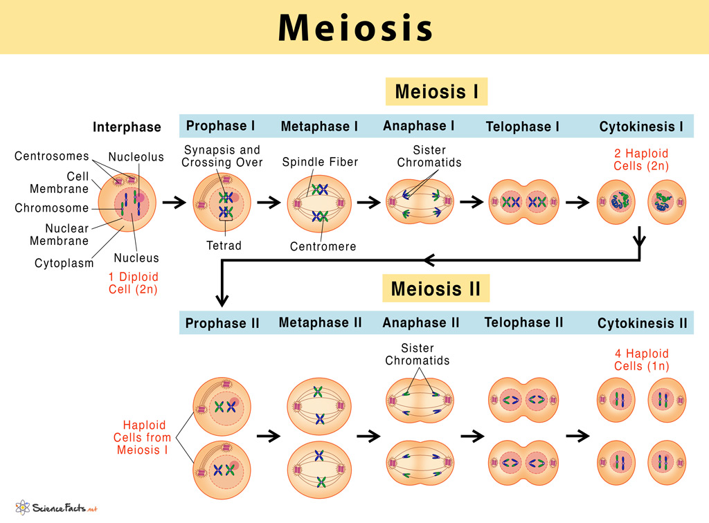
Meiosis involves two successive stages or phases of cell division, meiosis I and meiosis II. Each stage includes a period of nuclear division or karyokinesis and a cytoplasmic division or cytokinesis. Although not a part of meiosis, the cells before entering meiosis I undergo a compulsory growth period called interphase.
Interphase
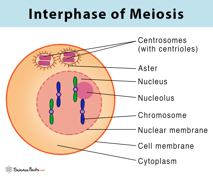
During this phase, the nuclear envelope remains intact. The chromosomes exist in the form of long, slender, and coiled chromatin fibers.
G1 phase: The first gap phase or the preparatory phase of cell division. During this phase, the cell increase in size by absorbing water from the cytoplasm and synthesize different types of RNA and proteins.
S phase: The period of DNA synthesis during which the genetic material present within the nucleus gets copied. Each chromosome duplicates to become two identical sister chromatids attached at a specific point, called the centromere. The centrioles get duplicated as well.
G2 phase: The second gap phase that happens after the DNA synthesis, but before prophase. During this phase, the cell continues to increase in size with the synthesis of RNA and proteins.
Steps of Meiosis I
1) Prophase I
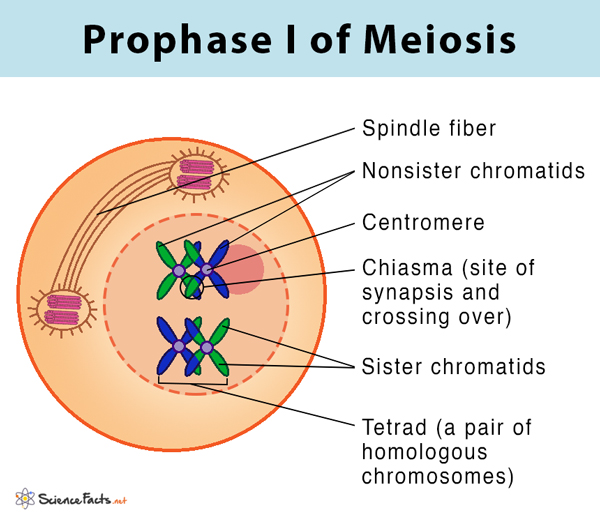
It is the longest phase of meiotic division involving a series of events and is divided into the following steps:
- The duplicated chromosomes condense, resembling an X-shaped structure with two sister chromatids that become distinctly visible within the nucleus.
- The homologous chromosome pair (one inherited from each parent) comes closer and associate along the entire chromosome length, forming a tetrad. Each tetrad is composed of four chromatids.
- The homologous chromosomes exchange parts of DNA with each other; this process is known as crossing over. The points of physical contact from which the genetic materials are exchanged are known as chiasmata.
- Spindle fibers originate from the centrioles on either side of the cell, getting attached to each chromosome’s centromere.
- The last step of prophase involves the breakdown of the nuclear envelope. The chromosomes then start moving towards the middle of the cell.
2) Metaphase I
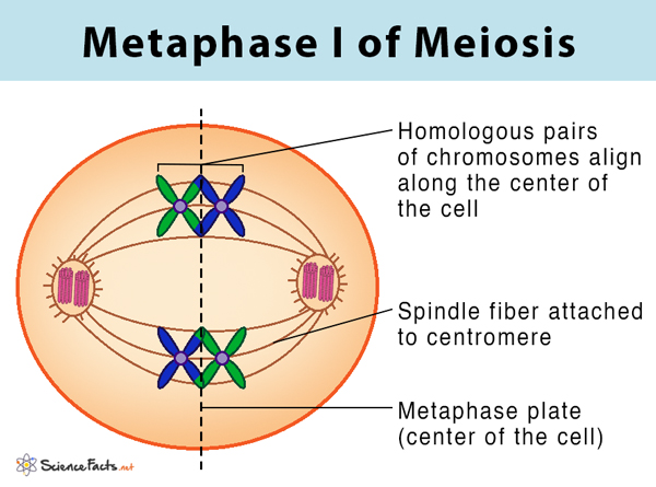
- Homologous chromosomes align along the center of the cell.
- The centrioles reach the opposite poles of the cell with the spindle fibers extending from them.
- The centromeres orient themselves towards the opposite poles of the cell.
3) Anaphase I
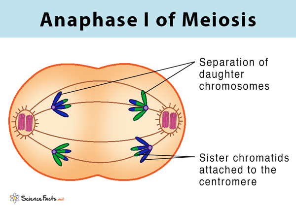
- The chromosomes with two sister chromatids are separated, and they begin to migrate to the opposite poles. This separation is achieved because of the contraction of the spindle fibers attached to each chromosome’s centromere.
- The homologous chromosomes start to migrate to the opposite poles.
4) Telophase I
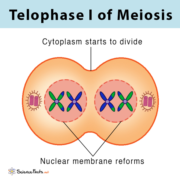
- The chromosomes stopmigrating with each pole containing a haploid number of chromosomes.
- The nuclear envelope is formed around the chromosome, and the spindle fibers disappear.
- The chromosomes uncoil and become less dense with the nucleolus appearing within the nucleus.
5) Cytokinesis I
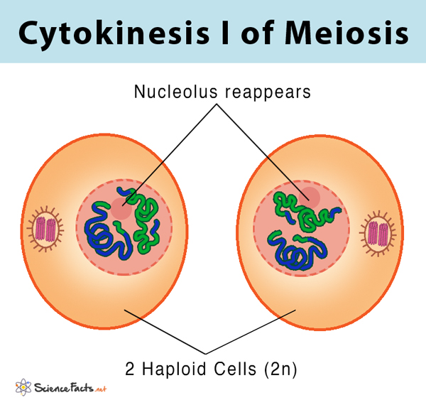
- It involves the division of the cytoplasm to produce two individual daughter cells. In animals, cytokinesis occurs by constriction of the cell membrane, while in plants, it happens through the formation of a cell plate. In most cells, cytokinesis occurs at the same time as telophase.
Result of Meiosis I
At the end of cytokinesis I, two different daughter cells are formed, each with half the number of chromosomes as the parent cell (having 23 chromosomes having 23 pairs of chromatids). Meiosis is thus also called the reduction division.
Steps of Meiosis II
The daughter cells produced in meiosis I enters the second round of division called meiosis II.
1) Prophase II
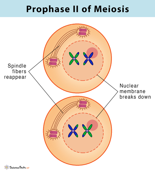
- The nuclear membrane initiates to break down, and the spindle fibers appear again.
- Each centriole divides, forming two pairs of centrioles.
- Chromosomes do not replicate any further in this phase of meiosis and begin migration towards the center of the cell.
2) Metaphase II
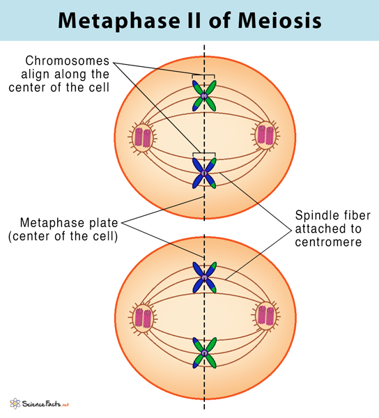
- Chromosomes arrange on the equator of the cell with the help of the spindle fibers.
- The centrioles are now at opposite poles in each of the daughter cells.
- Centromere divides, producing two sister chromatids, now known as daughter chromosomes, with the spindle fibers attached to each chromosome.
3) Anaphase II
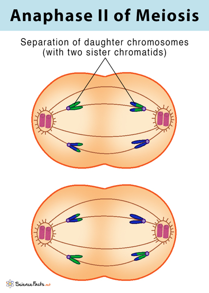
- The daughter chromosomes are pulled towards the opposite poles of the cells with the help of the spindle fibers. At the end of anaphase II, each end of the cell contains a complete set of chromosomes.
4) Telophase II
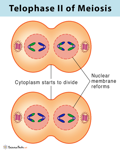
- The nuclear membrane forms around each chromosome with the disappearance of the spindle fibers.
- Nucleolus reappears as the cell prepares for the second round of cytoplasmic division.
5) Cytokinesis II
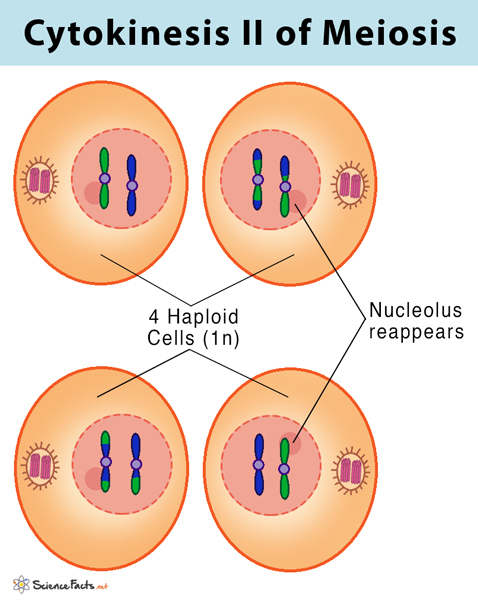
- This step is identical to cytokinesis I, involving the second cytoplasm division, resulting in the formation of two individual daughter cells.
What is the End Result of Meiosis
Thus at the end of meiosis II, four non-identical, haploid daughter cells are formed, each having half chromosome number as the original parent cell.
Main Differences Between Meiosis I and Meiosis II
In meiosis I, a pair of homologous chromosomes separate to produce two diploid daughter cells, each having half the number of chromosomes as the parent cell. In contrast, during meiosis II, sister chromatids separate to produce four haploid daughter cells. Also, unlike meiosis I, no genetic recombination by crossing over occurs in meiosis II.
Purpose of Meiosis
- Maintaining chromosome number in organisms: In humans, each cell typically contains 46 chromosomes organized into 23 pairs. To maintain the chromosome number generation after generation, the gametes formed from the meiotic division should contain half the number of chromosomes (23 chromosomes) as the parent cell. When the sex cells fuse to form a zygote, the usual chromosome number of 46 chromosomes is restored in the new individual. If the chromosomal reduction process is not maintained, it could cause genetic abnormality in the child.
- Creates genetic diversity: The exchange of genetic information between the pair of homologous chromosomes allows genetic variation among the population. These variations form the basis of the evolutionary process.
- Repairs genetic defects: The process of mixing chromosomes in meiosis, commonly known as recombination, helps repair genetic abnormalities in individuals produced through meiosis. When one of the parents has a genetic defect, recombination through meiosis can replace that abnormality in the next generation, allowing the formation of a healthy individual.
FAQs
Ans. Since meiosis occurs through the union of sex cells or gametes, it cannot happen asexually.
Ans. Non-disjunction is an error in meiosis when a pair of chromosomes has failed to separate at anaphase. In such cases, both the chromosome pair has passed on to the same daughter cell.
-
References
Article was last reviewed on Tuesday, May 16, 2023
Related articles
5 thoughts on “Meiosis”
Leave a comment
Popular Articles


Join our Newsletter
Fill your E-mail Address
cool
The information regarding the chromosome number of the two cells at the end of Meiosis I is incorrect. At the end of the first division, the cells are HAPLOID, not diploid; they are still double-stranded, but there is only half of the # that is in original cell.
1. Telophase I picture correctly states there is a haploid # of chromosomes at each pole… BUT THEN
2. Cytokinesis I picture mislabels them as two Diploid 2n cells.
3. Again correct in the “Results of Meiosis I” statement.
4. First diagram of the whole process is mislabeled at the end of Cytokinesis 1 and the Prophase II picture.
In the “script” is correctly states Meiosis is reduction/division, Meiosis I REDUCES the number of chromosomes, then Meiosis II DIVIDES them.
We have made the necessary changes
Cells after first division are not diploid.
They are haploid, with replicated chromosomes. They are no more “diploid” than any haploid cell after replication.
We have made the necessary changes