Spindle Fibers
What are Spindle Fibers
Spindle fibers are a network of filaments called microtubules that move chromosomes during eukaryotic cell division. Spindle fibers formed in mitosis are called the mitotic spindle, while those formed in meiosis are called the meiotic spindle. Microtubules that form the spindle fibers come from centrosomes.
Spindle fibers play a significant role during nuclear division. They are responsible for the separation of sister chromatids during mitotic and meiotic cell division.
When Do They First Become Visible
They begin to form during the prophase of cell division. Then, during the metaphase, the spindle fibers become visible as they radiate from the centrioles at the opposite poles.
Structure
What Are They Made Of
They are spindle-shaped fibers composed of 97% motor protein (tubulin) and 3% RNA. The microtubules are a polymer of ???? and ????-tubulin dimers.
Spindle fibers, associated proteins, condensed chromosomes, and centrosomes or asters together form the spindle apparatus.
Its Formations
At the beginning of the nuclear division, two wheel-shaped organelles called centrioles position themselves at opposite ends of the cell, forming two cell poles. On getting signals for cell division, long protein fibers called microtubules extend from the centrioles in all directions, forming spindle.
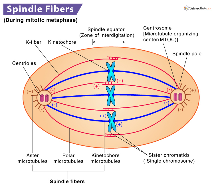
In mitosis, these filaments form at opposite poles of the cell and meet at the equatorial plane. Together they produce a spindle-shaped structure, which is widest at the middle and tapered towards both ends. These are called spindle fibers.
Types of Mitotic Spindle
There are three types of mitotic spindle based on their composition. They are named and discussed below.
- Kinetochore Microtubules: Kinetochores are macromolecular complexes assembled on each chromosome’s centromere during mitosis. They act as a link between DNA and microtubules. Kinetochore microtubules attach the chromosomes to the spindle pole and help pull apart the sister chromatids at the time of cell division.
- Aster Microtubules: They are radial microtubular assemblages found in animal cells that anchor the spindle poles to the cell membrane. They ensure correct positioning and orientation of the mitotic spindle apparatus based on cell polarity. They also determine the region of the cleavage furrow that splits the dividing cell in half during cytokinesis.
- Polar Microtubules: Spindle microtubules that do not attach to the chromosomes are called polar microtubules. They overlap at the spindle midzone and push the spindle poles apart via motor proteins, contributing to cell elongation.
Functions
Role in Mitosis
Spindle fibers play a significant role in mitosis. They remain highly active during this phase and equally divide the chromosomes in a parental cell into two daughter cells. Let us discuss the role of spindle fibers in each step of mitosis.
Prophase
Spindle fibers start to form the centrioles at the opposite poles of the cell. In animal cells, the mitotic spindle appears as asters that surround each centriole pair. The sister chromatids attach to spindle fibers at their kinetochores during this phase. Also, the cell elongates as spindle fibers stretch from each pole.
Metaphase
As the mitotic spindle extends from each pole, they create tension. As a result, chromosomes line up at the metaphase plate. Microtubules from opposite spindle poles get attach to the two sister chromatids of each chromosome. In metaphase, the spindle has attached all the chromosomes and arranged them at the middle of the cell, making it ready for division.
Anaphase
As the spindle fibers attach to a protein complex called kinetochores, they either generate or transduce force to segregate and move sister chromatids to their opposite poles. Interpolar microtubules help in the lengthening and elongation of the cell to provide room for cell division.
Telophase
During this phase, the separated sister chromatids (now known as daughter chromosomes) reach the opposite poles and get enclosed within two new nuclei. The spindle fibers are found to disappear.
Cytokinesis
The cytoplasm divides, and two new daughter cells are formed with an equal number of chromosomes. Spindle fibers ensure this process.
Chromosome Movement
Cell movement occurs when microtubules of spindle fibers interact with motor proteins. Motor proteins like dyneins and kinesins move along microtubules causing the spindle fibers to contract (shorten) or relax (elongate). The disassembly and reassembly of microtubules produce the chromosomal movement required for cell division.
Spindle fibers help in chromosomal movement during cell division by attaching to centromeres (the primary constriction site of a chromosome). Kinetochores produce fibers that attach sister chromatids to the spindle fibers. Both the kinetochore and polar microtubules work together on segregating chromosomes. Spindle fibers that do not bind chromosomes during cell division extend from one cell pole to the other. These fibers overlap and push cell poles away from one another resulting in cell elongation, preparing the cell for cytokinesis.
-
References
Article was last reviewed on Friday, February 17, 2023
Related articles
Popular Articles


Join our Newsletter
Fill your E-mail Address