Centriole: Definition, Structure, and Functions
What are Centrioles
Centrioles are cylindrical organelles composed of a microtubule protein called tubulin. They are found in all eukaryotic cells.
Eukaryotes contain a mother centriole, and a daughter centriole bound together and arranged near the nucleus at right angles. In a cell, they aid in cell division by facilitating the separation of chromosomes, containing DNA, the genetic material of the cell. Centrioles are absent in prokaryotes, red algae, yeast, cone-bearing, and flowering plants, and some non-flagellated or non-ciliated protozoans such as amoebae.
Location
They are found in all animal cells and a few lower plant species. Within a eukaryotic cell, centrioles are present close to the nucleus. During mitotic division, they form a spindle of microtubules called the mitotic apparatus that moves towards the opposite ends of the nucleus. When a centriole bears a flagellum or cilium attached to the mitotic apparatus, it is called the basal body.
Structure and Characteristics
Size and Shape
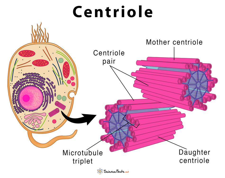
A centriole is a small set of microtubules arranged in the form of an open-ended cylinder with a length of 0.3–0.7 um and a diameter of 0.15–0.25 um. Centrioles are visible under a light microscope but can be viewed in detail only under an electron microscope. A pair of centrioles lies in a common specialized part of the cytoplasm called centrosphere, which is devoid of any other organelle. The complex, formed together with centrioles and centrosphere, is called the centrosome.
Composition: What are Centrioles Made Of
A centriole is made of nine sets of short microtubule triplets arranged in a cylinder with no central microtubules, making it a hollow center, this arrangement is thus called 9 + 0.
Each triplet microtubule consists of a complete microtubule, the A-tubule, on which two additional partial microtubules, the B- and the C-tubules, are assembled. The adjacent microtubules are connected by С-A protein bonds, while the center contains a rod-shaped mass made of proteins, known as the hub. From the hub, develops nine protein strands towards the peripheral triplet microtubules called spokes. This makes the centriole looks like a cartwheel.
Deviations from this arrangement are found in Brachyura species, embryos of Drosophila melanogaster, and sperm cells of Caenorhabditis elegans.
Functions: What Does the Centriole Do In a Cell
Main Functions
Centrioles serve as the major microtubule-organizing centers, which is an important event in two major cellular processes:
- Cell Division (allows chromosomes to move inside the cell): The centrioles play active roles in almost every phase of cell division. It helps in the formation of aster and spindle fibers and attaching them to the chromosomes. This helps in equal distribution of the genetic material among daughter cells. Thus a human cell without a centriole cannot divide.
- Formation of Cilia and Flagella (to provide the cell with the ability to sense extracellular signals and also helps in movement): Occurs by a process known as ciliogenesis, centrioles give rise to basal bodies, with both having an identical size, structure, and chemical compositions. Basal bodies later develop into cilia and flagella.
Other Important Roles
- Centriole Duplication: Forms new centrioles that divide and get duplicated during S phase of the cell cycle.
- Receives and Processes External Stimuli: Helps to determine the beating pattern of cilia and flagella, and thus influence the ability to receive external stimuli.
- Cellular Organization: Helps in the spacial arrangement and positioning of other organelles. This is important for the proper and coordinated functioning of the cell.
- Intracellular Transport: Allows transport of signaling molecules and other substances within the cell due to the beating of cilia and flagella.
- Animal Growth and Development: Maintains proper symmetry during embryonic development in mammals.
- Fertility: Helps in the formation of sperm flagellum and in sperm movement. Centrioles are also responsible for the development of the embryo after fertilization.
Centriole vs Centrosome: What Are the Differences Between a Centriole and a Centrosome
| Basis | Centriole | Centrosome |
| Structure and Composition | Cylindrical shaped that are composed of globular protein called tubulin | Have no definite shape and are comprised of centrioles as well as the dense protein called peri-centriolar material |
| Location | Found near the nucleus | Found in the specialized DNA sequence of a chromosome that links a pair of sister chromatids |
| Number | Seen in pairs of two during cell division. After division, each daughter cell has one pair of centrioles. | Seen as a single pair located near the nuclear membrane at the start of the cell division. After division, each daughter cell receives one copy of a centrosome. |
| Division | Replicate in the S phase and two copies of a pair of centrioles are formed | Replicate in the S phase but produce only a single copy of themselves |
| Main Functions | Forming mitotic spindle and aster during cell division. The centrioles also help in the formation of cilia and flagella that aid in movement of the sperm and ova during fertilization | Organizing the centrioles and their microtubules during cell division. The centrosome helps to maintain chromosome number in daughter cells |
FAQs
Ans. Edouard Van Beneden made the first observation of centrosomes in 1883, which is found to be composed of two orthogonal centrioles. In 1895, Theodor Boveri named the organelle a ‘centrosome’.
Ans. All prokaryotic organism including bacteria do not have membrane-bound organelles including centrioles.
Ans. Although plants do not have centrioles, they are still able to form mitotic spindle from their centrosome region just outside the nucleus, which helps in their cell division.
Ans. The structure that is produced when protein fibers radiate from centrioles is called aster.
-
References
Article was last reviewed on Wednesday, September 2, 2020
Related articles
Popular Articles


Join our Newsletter
Fill your E-mail Address