Pinocytosis
What is Pinocytosis
Pinocytosis, also known as fluid endocytosis, fluid-phase endocytosis, and bulk-phase pinocytosis, is defined as the process where a cell ingests tiny particles suspended in the extracellular fluid by forming an endocytic vesicle called a pinosome.
The term pinocytosis combines two Greek words, where ‘pino’ means ‘to drink’, and ‘kytos’ denotes ‘cell’. So, the word stands for ‘cell drinking’. It involves the transport of extracellular fluids along with its contents of small dissolved molecules (solutes). That is why pinocytosis is considered a form of endocytosis.
Is Pinocytosis a Form of Active or Passive Transport
Pinocytosis requires a considerable investment of cellular energy in the form of ATP to run. Therefore, it is an active transport mechanism.
Though it is not specific, certain ions or amino acids can trigger the process.
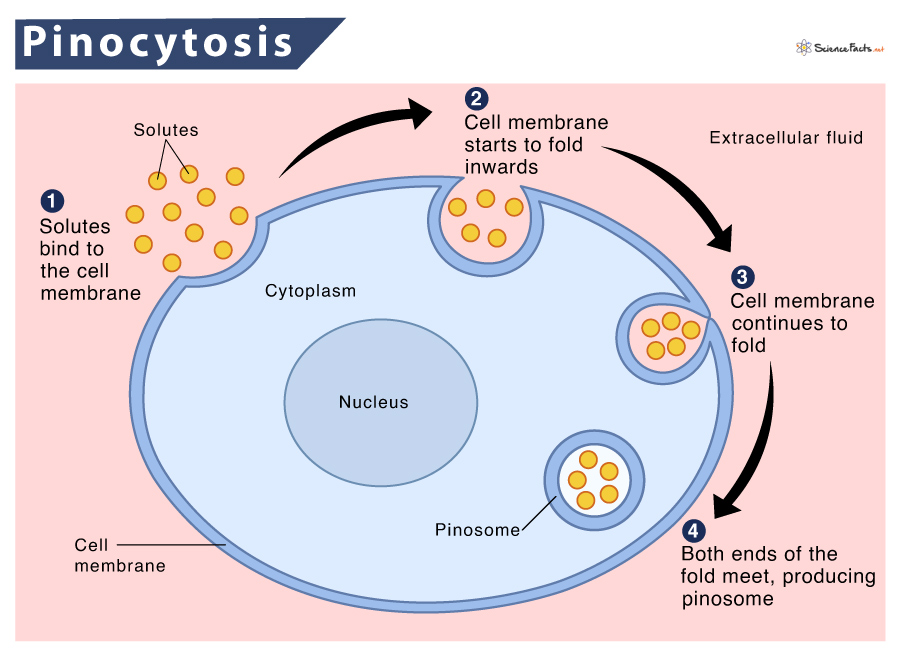
What Happens During Pinocytosis
During pinocytosis, the cell ‘drinks’, i.e., it takes in the surrounding fluids along with the solutes present in it. So, this process can be recognized when the cell is engulfing extracellular fluid (ECF).
Various molecules, such as ions, sugars, and amino acids, activate the process when they contact the plasma membrane. This phenomenon induces the development of small pockets (fold) on the membrane around ECF (and minute dissolved solutes), and the consequent internalization of these materials occurs inside the cell.
Steps of the Process
The basic steps of pinocytosis are described below:
Step 1 – Initiation: In the initial stage, an inducer molecule, such as sugar, protein, or ion, comes in contact with the plasma membrane. As a result, an ionic interaction occurs between the positively charged inducer and the negatively charged cell membrane. This phenomenon triggers the binding of the molecule to the membrane.
Step 2 – Folding of the Membrane: Following binding, the cell membrane gets stimulated to fold inwards and form a small open-ended pocket, or invagination, around the fluid containing the molecule.
Step 3 – Invagination and Engulfment of the Fluid: The cell membrane continues to fold inwards with the fluid and dissolved solutes in small pockets. Later, they start to reconnect at the open end of the invagination to enclose the fluid and solutes.
Step 4 – Detachment of the Pocket: This is the final stage when both ends of the invagination meet. It gives rise to a vesicular structure called a pinosome that contains extracellular fluid and dissolved solutes. Afterward, this pouch gets detached or pinches off from the cell membrane. The molecules present inside the vesicle are eventually released to be used by other parts of the cell, thus completing the process.
Types
Pinocytosis is classified mainly depending on the size of the transferred molecules or based on the method of vesicle formation.
Based on the Size of the Molecule
Depending on the size of the molecules to be transferred, pinocytosis has two broad divisions:
Micropinocytosis
Going by the name, it refers to the uptake and internalization of small molecules. In this case, the vesicle or micropinosome formed measures around 0.1um in diameter. As a result, tiny indentations develop on the cell membrane.
An example of this phenomenon is Caveolin-mediated pinocytosis (has been described later), formed in the epithelial cells of blood vessels.
Macropinocytosis
Cells use this process to draw larger materials from the extracellular environment inside the cell. So, in this case, the vesicle or the macropinosomes are large with a diameter of around 0.5-5 µm.
Macropinocytosis is non-specific and thus involves the uptake of a variety of materials ranging from antigens to nutrients.
This type of pinocytosis helps immune cells to detect soluble antigens, among other materials in ECF. As soon as the inducers trigger the process, the cell’s cytoskeleton rearranges the actin filaments in the cell membrane to produce protrusions or ruffles to form macropinosomes. The vesicle matures in the cytoplasm. Later, they either fuse with the lysosomes or migrate towards the cell membrane for recycling.
This process is commonly seen in white blood cells (WBCs) like macrophages and dendritic cells.
Based on the Receptor and Their Mode of Action
Depending on the receptor involved in the process and the mechanism of vesicle formation, pinocytosis further has four divisions. Among the four types, one of them is macropinocytosis, which is described in the previous section. The rest three divisions are as follows:
Clathrin-Mediated Pinocytosis
Here, the cells take up a wide range of molecules, including proteins, hormones, and various metabolites. Like macropinocytosis, the cell membrane undergoes a conformational change following the attachment of molecules to the surface receptors.
This receptor-mediated pinocytosis is the best form of endocytosis to uptake selective macromolecules. Initially, these molecules adhere to specific cell surface receptors known as clathrin-coated pits. Later the pits bud through the membrane to develop tiny clathrin-coated vesicles, which contain the receptors and their bound macromolecules. Finally, the vesicles fuse with early endosomes. They are then driven for transportation to the lysosomes or recycled to the plasma membrane.
Caveolae-Mediated Pinocytosis
It helps in the transport of small materials (solutes and molecules) inside the cell. It is an example of micropinocytosis. The adipocytes (fat cells) and endothelial cells of blood vessels exhibit this phenomenon.
Caveolae are 50–80 nm cup-shaped pits usually found immobile in the plasma membrane. They are often found associated with two classes of proteins, caveolins, and cavins. When a caveolae-liked receptor is activated, it can bud off from the cell membrane to form vesicles. Various solutes or molecules present in the extracellular fluid interact with the caveolae complex and caveolin proteins on the membrane. As a result, the internalization of the complex and proteins take place.
Clathrin and Caveolae-Independent Pinocytosis
This type of pinocytosis does not depend on receptors or other coat proteins or material stimuli to develop vesicles. Due to this, it allows the transportation of a wide variety of materials, including various pathogens, receptors, and toxins.
Actin and associated proteins play an essential role in the formation of vesicles.
Functions
- Being a form of active transport, it plays a significant role in several cellular processes like the uptake of nutrients, excretion of waste materials, and signal transduction.
- Various unicellular organisms like amoeba and paramecium use this process to uptake nutrients (certain sugars, amino acids, organic acids, and many inorganic ions).
- Aids in the bulk transport of dissolved molecules like fats and vitamins in higher organisms, like humans.
- Being non-specific, it allows absorption and transport of many different molecules at once.
- Excretion of waste materials from the cells, such as removing water and waste products from the kidney.
- The immune system cells, such as the macrophages and dendritic cells, use it to detect antigens in the extracellular fluid.
Examples
- In the gastrointestinal(GI) tract: Absorption of nutrients in the microvilli of the small intestine from the GI tract.
- In egg cells: Absorption of nutrients by egg or ovum of the female reproductive system from mucosa cells of the fallopian tube before fertilization.
- In renal tubular cells: Reabsorption of essential nutrients and excess water in the kidney tubules during urine formation.
- In immune cells: Detection of the presence of antigens in ECF.
- In unicellular organisms: Uptaking of water and dissolved nutrients by single-celled organisms like amoeba and paramecium.
- Cell maintenance: Recycling the cell membrane components and maintaining their size by most body cells.
Pinocytosis vs. Phagocytosis
As discussed in our ‘endocytosis’ article, there are four types of endocytosis. Among them, two are – pinocytosis and phagocytosis.
Though both of them are a variation of endocytosis, they have contrasting work mechanisms.
What is the Difference between Pinocytosis and Phagocytosis
| Basis | Pinocytosis | Phagocytosis |
| 1. The Process | It is a type of endocytosis by which the cell ingests solid particles sized 0.5 µm or less. | It is a type of endocytosis by which the cell ingests solid particles greater than 0.5 µm. |
| 2. Mechanism of Intake | Intake molecules by invagination and forming a pocket-like structure in the cell membrane. | Intake molecules by forming pseudopodia (false feet). |
| 3. Nature of Ingested Particles | The ingested particles are dissolved in liquid. | The ingested particles are solid. |
| 4. Substrate Specificity | Non-specific and transports a large number of molecules at once. | Specific and only transports a particular molecule at once. |
| 5. Energy Utilization | The energy requirement is low compared to phagocytosis, i.e., lesser ATPs are needed. | The energy requirement is high compared to pinocytosis, i.e., more ATPs are needed. |
| 6. Vesicles Formed | Vesicles formed are called pinosomes. | Vesicles formed are called phagosomes. |
| 7. Main Purpose | To intake necessary nutrients. | To engulf foreign invaders. |
| 8. Involvement of Lysosome | Pinosome does not fuse with a lysosome. | Phagosome fuses with a lysosome, forming phagolysosome or food vacuole. |
| 9. Types of Particles Ingested | Molecules like sugar, amino acids, vitamins, and ions are taken in. | Bacteria, viruses, and any other foreign materials are ingested. |
| 10. Site of the Process | Found in almost all cells, including the secretory cells and epithelial cells of the blood vessels. | Found in immune cells like macrophages and neutrophils. |
| 11. Spontaneity | It is a constitutive process, which occurs continuously. | It is a triggered process, which occurs occasionally. |
| 12. Mode of Digestion | The cell engulfs food that is already broken down or digested. | The cell engulfs whole particles, which later get broken down or digested by hydrolytic enzymes. |
Similarities
Despite numerous differences, they share some similarities. They are given below:
- Types of endocytosis. So, they help to draw extracellular materials inside the cell.
- Involve the folding of the plasma membrane to form a vesicle or pouch-like structure.
- Require energy (ATP) for cellular transport.
FAQs
Ans. Pinocytosis refers to the ingestion of liquid (water) into a cell by the budding of small vesicles from the cell membrane. On the contrary, receptor-mediated endocytosis refers to an endocytotic mechanism in which specific molecules (solutes) are ingested into the cell by developing endosomes via special cell receptors. Pinocytosis is a less efficient form of endocytosis than receptor-mediated endocytosis.
Ans. Both pinocytosis and receptor-mediated are types of endocytosis. Both use the cell membrane to uptake proteins and other substances that are not easily diffusible into the cell through the plasma membrane. They are categorized under active transport as they require ATP.
-
References
Article was last reviewed on Friday, February 17, 2023
Related articles
Popular Articles


Join our Newsletter
Fill your E-mail Address