Animal Cell: Parts and Structure with Functions
What is an Animal Cell
An animal cell is defined as the basic structural and functional unit of life in organisms of the kingdom Animalia. They have a distinct nucleus with all cellular organelles enclosed in a membrane, and thus called a eukaryotic cell.
Structure and Characteristics of an Animal Cell
The shape of a typical animal cell varies widely from being flat, oval to rod-shaped, while others assume shapes such as curved, spherical, concave, and rectangular. An animal cell ranges in size from 10 to 30 µm.
Under the microscope, an animal cell shows many different parts called organelles, that work together to keep the cell functional.
Parts of an Animal Cell
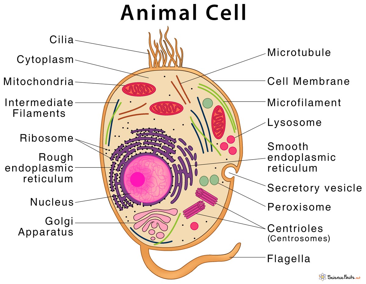
1) Centrioles
They are paired tube-like organelle composed of a protein called tubulin. Centrioles are about 500nm long and 200nm in width that are found close to the nucleus and helps in cell division. They are also found in cilia and flagella.
Functions
- Helping in cell division by allowing separation of chromosomes
- Helping in cell movement
2) Centrosomes
Also known as the ‘microtubule-organizing center‘ of the animal cells, they are made up of two centrioles, linked together by interconnecting fibers. The centrosome is similar to DNA, where one centrosome from each parent cell is transferred to the daughter cell.
Functions
- Helping in the separation of chromosomes during cell division
- Maintaining the chromosome number during cell division
- Organizing microtubules and thus providing cell shape
3) Lysosomes
They are small membrane-bound organelles, filled with hydrolytic enzymes that can break down biomolecules such as carbohydrates, fats, and proteins. The size of lysosomes varies among different cell types, with the largest ones measuring more than 1.2 μm.
Functions
- Digesting complex biomolecules such as carbohydrates, lipids, proteins, and nucleic acids
- Destroying the organelles that are not functioning properly
- Removing cellular waste products from the cell
4) Cell Membrane or Plasma Membrane
It is the outermost membrane of an animal cell having a thickness of 5-10 nm that separates the interior of the cell from outside. The cell membrane is selectively permeable in nature, consisting of a lipid bilayer with proteins, glycolipids, and cholesterol attached to them in a specific pattern.
Functions
- Protecting the integrity of the cell from the outside environment
- Allowing selective entry and exit of substances in and out of the cell
- Maintaining the shape of the cell
- Providing mechanical support to the cell
- Keeping the cell turgid and helping in their growth
- Helping in cellular communication
5) Endoplasmic Reticulum (ER)
It is a continuous membrane-bound organelle, that remains distributed throughout the cytoplasm and forming connections between nuclear envelope and the cell membrane. There are two types of ER: rough endoplasmic reticulum (RER) and smooth endoplasmic reticulum (SER). The surface of RER is studded with ribosomes, which gives it a rough appearance, while SER is devoid of ribosomes.
Functions
- Helping in protein synthesis (RER)
- Synthesizing essential lipids such as phospholipids and cholesterol (SER)
- Producing steroid hormones and helping in their secretion (SER)
- Helping in the metabolism of carbohydrates (SER)
- Helping in the maturation of proteins (RER)
6) Golgi Apparatus
Also known as the Golgi body or Golgi complex, it is a series of five to eight cup-shaped, membrane-covered sacs called cisternae. They receive proteins and lipids from RER, which are then modified, sorted, packaged, and transported to their destination.
Functions
- Processing, packaging and transporting or secretion of the proteins to their target organs
- Performing protein modifications such as phosphorylation and glycosylation
- Breaking down of proteins into smaller fragments
7) Microfilaments or Actin filament
They are a network of rod-shaped proteins called actin that forms a part of the cell cytoskeleton. Microfilaments are the thinnest of all the cytoskeletal filaments, having a diameter of about 6-7 nm.
Functions
- Helping in the contraction of muscles
- Causing cell movement
- Allowing transport of nutrients, waste products, and cell organelles from one part of the cell to another (cytoplasmic streaming)
- Helping in cell division
8) Microtubules
They are hollow tubes composed of the protein tubulin. They are the largest of all cytoskeletal filaments, measuring about 24 nm in thickness.
Functions
- Forming an important component of cilia and flagella that helps in cell movement
- Helping in cell division
- Helping in the movement of nutrients, organelles, and waste products throughout the cell (cytoplasmic streaming)
- Helping in cellular communication
9) Intermediate Filaments
They are elongated fibrous proteins forming a coiled-coil structure. Intermediate filaments have a diameter of 8-10 nm that are intermediate in size compared to the other two cytoskeletal elements. Microtubules, together with microfilaments and intermediate filaments, complete the cytoskeleton of the cell.
Functions
- Providing structural and mechanical support to the cell
- Maintaining cell shape
- Helping in the movement of cell organelles and nutrients within the cell (cytoplasmic streaming)
10) Mitochondria
They are double-membrane-bound organelle with a size of 1 – 10 microns, that can be spherical or rod-shaped. Mitochondria are commonly called the ‘Powerhouse of the cell‘, producing ATP, the energy currency that drives all cell-based metabolic activities.
Functions
- Promoting the growth of new cells and in cell multiplication
- Controlling various cellular activities like respiration, metabolism, cell division, and cell death
- Maintaining an adequate concentration of calcium ions within the cell
- Playing an essential role in apoptosis or programmed cell death
11) Nucleus
It is a spherical double membrane-bound cell organelle that contains the genetic material of the cell. They are the largest and most prominent of all cell organelle. A nucleus has four main parts:
Nuclear membrane or Nuclear envelope: A double-membrane structure that separates cytoplasm from the nucleus
Chromatin threads or Chromosomes: Genetic material of the cell
Nuclear sap or Nucleoplasm: A Clear transparent liquid that contains chromosome
Nucleolus: A membrane-less structure that produces ribosome
Functions
- Controlling the activities of the entire cell
- Storing the genetic material of the cell
- Performing cell growth and reproduction
12) Peroxisomes
They are single membrane-bound cell organelle with a size of 0.1-1 mm that contains a range of digestive and oxidative enzymes. Peroxisomes vary in shape, size, and number, depending upon the energy requirements of the cell.
Functions
- Breaking down of fatty acids to provide energy to the cell
- Performing lipid synthesis
- Detoxification of alcohols and other toxic compounds
13) Ribosome
They are minute particles present in large numbers, either found attached to the endoplasmic reticulum or remain free in the cytosol. Ribosomes are the protein-synthesizing center of the cell.
Functions
- Producing proteins required for all cellular activities including growth, metabolism and cell division
14) Cytoplasm
They are the colorless, semifluid substance of a cell that covers the entire space except the area enclosed by cell organelles. The cytoplasm is composed of about 80% water and the rest are organic and inorganic compounds.
Functions
- Acting as the site of various cellular activities such as respiration, cell division and elimination of waste products
- Providing raw materials necessary for the chemical reactions within the cell
- Maintaining turgidity of the cell thus keeping cell shape
- Keeping the cell organelles in position
15) Cilia and Flagella
They are fine, hair-like projections that extend from the body of many cells and are made of microtubules. Cilia and flagella vary in terms of length and numbers based on the types and functions of the cell.
Functions
- Helping in cell movement
- Allowing to sense changes in the environment
- Helping in the movement cell organelles, nutrients, and waste products inside the cell (cytoplasmic streaming)
Most Common Types of Animal Cells
1) Skin Cells: Forms the external barrier of our body that provides protection. Skin cells are of two types -keratinocytes and melanocytes.
2) Muscle Cells: Present below the skin cell, they help in body movement. Muscle cells are of three types – skeletal muscle cells, cardiac muscle cells, and smooth muscle cells.
3) Blood Cells: Found mainly in the blood, they can be divided into two types – red blood cell (RBCs) and white blood cell (WBCs).
4) Nerve Cells: Basic units of the nervous system. They receive, carry, and deliver signals throughout the body.
5) Fat Cells: Also known as adipocytes or lipocytes, they are used to store fats and other lipids as energy reserves.
FAQs
Ans. Animal cells have centrioles, centrosomes, and lysosomes while plant cells do not have any of them.
Ans. Sometimes, the digestive enzymes present in the lysosomes end up damaging the lysosome itself, and this can ultimately cause the destruction of the cell (autolysis). Hence, lysosomes are also called the suicidal bags of the cell.
References
-
References
Article was last reviewed on Thursday, February 2, 2023
Related articles
3 thoughts on “Animal Cell: Parts and Structure with Functions”
Leave a comment
Popular Articles


Join our Newsletter
Fill your E-mail Address
Related Worksheets
This is cool! Thanks whoever did this!
This is amazing
Thanks so much