Cell Junctions
What is Cell Junction
Cell junctions, alternatively known as intercellular bridges, are specialized protein structures that provide a physical connection between the adjacent cells or between a cell and the extracellular matrix in plants and animals. These junctions are only found between immobile cells and are especially abundant in epithelial tissues.
As they consist of cell adhesion molecules, they hold cells together. Cell junctions also enable cellular communication, provide tissue structure, and transport material between cells.
What are the Different Types of Cell Junctions
The ways of communication in plants and animal cells are different from each other. However, in each case, they communicate between their neighboring cells via intercellular junctions, which are specialized regions of contact between the cell membranes of adjacent cells. The different types of cell junctions are listed below:
In Plants
- Plasmodesmata
In Animals
Three major types of junctions are unique to animals. They are:
- Tight Junctions (Occluding Junctions or Zonula Occludens)
- Gap Junctions (Septate Junction)
- Anchoring Junctions
In addition to all the above-mentioned intercellular junctions, the extracellular matrix in animal cells also helps in cell adhesion and communication through conformational changes.
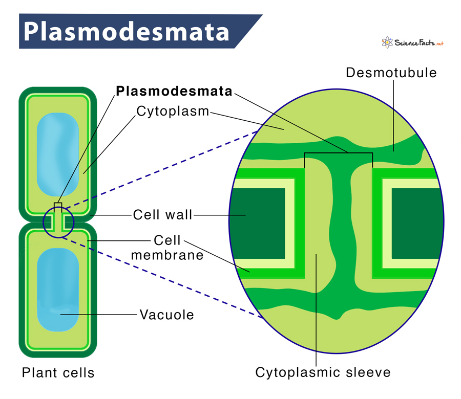
Plasmodesmata
As plant cells remain separated by the cell wall, they have developed some structural modifications called plasmodesmata. These are numerous channels passing between the cell walls of adjacent plant cells, connecting their cytoplasm.
Location
As mentioned, plasmodesmata are present between the two neighboring plant cells.
Structure and Composition
Plasmodesmata is a tube-like structure linking one cell to another. A membrane surrounds the open space of the tube and the desmotubule, containing tightly packed endoplasmic reticulum (ER), runs in the center. The ER lies continuously between the two cells. The cytoplasmic sleeve runs between the desmotubule and the membrane, where most transfer of molecules occurs. The sleeve contains various structures, such as actin and myosin, which provide contractile forces to transport.
Functions
- They are active cell components in intercellular transport, during development, and in the mature tissue.
- They facilitate the movement of molecules between cells, ranging from small photosynthetic products to large proteins and mRNA.
- They link tissue cells to one another, thus helping in tissue growth and development.
- They offer direct contact with adjacent cells.
- They help regulate the function of sieve-tube cells by the companion cells in the phloem through symplastic transport.
Tight Junctions
Tight junctions (TJs), also called zonula occludens or occluding junctions, are selectively permeable seals in the body’s internal and external surfaces. The term ‘zonula’ means small zone or encircling belt, while ‘occludens’ roots from the Latin word ‘occludere,’ meaning ‘to close up.’ Thus TJs create a small zone occluding the extracellular space, binding two cells together into a leakproof sheet.
Location
Tight junctions are located at the border between apical and lateral membranes between epithelial cells.
Structure and Composition
TJs are composed of numerous essential proteins, including:
- Claudins – form the backbone of tight junction strands
- Occludins – maintain the barrier between adjacent cells
- Junctional adhesion molecules (JAMs) – immunoglobulin (antibody) proteins that help seal the intercellular space between two cells
- Zonula occludens (ZO) – link the tight junction to each cell’s cytoskeleton
Among all, the occludins and claudins are the primary components of tight junction strands. When fully formed, a TJ is not one long, continuous seal. Instead, it appears like a series of local seals joined together in a maze-like fashion.
Functions
- They serve as a structural support mechanism and keep the adjacent epithelium together.
- They demarcate the apical region from the basolateral region. Thereby, TJs serve as a physical barrier within the membrane, thus establishing cell polarity.
- Their complexes form paracellular channels, allowing the selective diffusion of ions and solutes through the intercellular space.
- Their functions depend on the type of epithelium in which they reside. For instance, they help prevent the leakage of digestive enzymes into our bloodstream in the digestive system. On the other hand, they keep us somewhat waterproof in the skin and help keep allergens out of our bodies.
Gap Junctions
Gap junctions, also known as communicating junctions, macula communicans, or nexuses, are connections that form bridges the cytoplasm of two adjacent cells directly. These junctions consist of several transmembrane channels called pores, found in a closely packed arrangement. The number of gap junctions presents between two cells varies from one cell to another.
Location
Gap junctions occur throughout the body, including epithelia or body coverings, nerves, cardiac muscle, and smooth muscle.
Structure and Composition
Each channel comprises two half-channel (hemichannels) present in each cell’s membrane. These half channels join together, bridging the extracellular space and thus forming the entire channel that spans both cell membranes.
Each of these halves is called a connexon. Each connexon is composed of six symmetrical integral membrane protein units called connexins. Together, each channel is made up of 12 circularly arranged protein units.
Functions
- Their primary role is to coordinate the activity of adjacent cells. For instance, when heart cells need to beat in unison, gap junctions transmit electrical signals.
- They allow direct passage of molecules between two cells. The molecules that may cross this channel include ions, regulatory proteins, and metabolites.
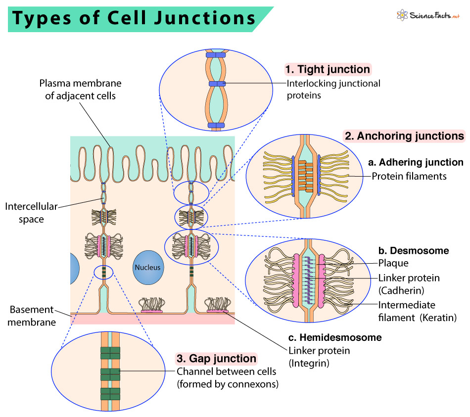
Anchoring Junctions
Anchoring junctions are cell junctions that remain anchored to adjacent cells while being attached to components of the extracellular matrix.
Location
They are commonly found in tissues prone to constant mechanical stress, e.g., skin and heart.
Structure and Composition
Anchoring junctions are composed of several transmembrane linkers like cadherin and integrins. They also use intermediate filaments and actin filaments as the cytoskeletal anchor.
Functions
- They play an essential role in keeping the cells together and in the structural cohesion of tissues.
- They stabilize the cell’s position, provide stability and rigidity, and support tissue integrity by holding cells together.
- These junctions also form a tight seal between neighboring cells to restrict the flow of molecules between cells and from one side of the tissue to the other.
- They also regulate the motility of both single cells and cellular masses through their substrates.
There are three types of anchoring junctions. They are desmosomes, hemidesmosomes, and adherens junctions.
a) Desmosomes
Desmosomes are protein attachments between adjacent cells. It is found only in animal cells.
Inside the plasma membrane, a desmosome bears a disk‐shaped structure from which protein fibers extend into the cytoplasm. Some of these proteins extend across the membrane, while others anchor the junction within the cell.
Specialized adhesion proteins, called cadherins, are found on the membranes of both cells and interact in the space between them, holding the membranes together. Inside the cell, the cadherins attach to a cytoplasmic plaque, which connects to the intermediate filaments and helps anchor the junction.
Desmosomes act like spot welds to hold together tissues that undergo considerable stress, such as skin or heart muscle. They pin adjacent cells together, ensuring that cells in organs and tissues that stretch, such as skin and cardiac muscle, remain connected in an unbroken sheet.
b) Hemidesmosome
These structures look similar to the previous type but are different functionally and in their content. Like desmosomes, they also connect the basal surface of epithelial cells via intermediate filaments to the underlying basal lamina. However, unlike desmosomes, their transmembrane proteins are integrin.
c) Adhering Junctions
In adherens junctions, the cytoplasmic actin filaments act as the cytoskeletal anchor. Like desmosomes and hemidesmosomes, their transmembrane anchors are composed of cadherins and integrins. Cadherins anchor to other cells, and integrins anchor to the extracellular matrix.
Extracellular Matrix of Animal Cells
Animal cells sometimes release materials into the extracellular space, most of which contain collagen protein. These collagen fibers are interwoven with carbohydrate-containing protein molecules called proteoglycans, collectively called the extracellular matrix.
Cells possess protein receptors on the extracellular surfaces of their cell membranes. When a molecule within the matrix gets bind to the receptor, it changes the structural conformation of the receptor. The receptor, in turn, changes the structure of the microfilaments positioned immediately inside the cell membrane. As a result of these conformational changes, some chemical signals inside the cell get induced. Upon reaching the nucleus, the signals switch on or off the transcription of DNA, affecting the production of associated proteins.
FAQs
Ans. Gap junction is unique to animals.
Ans. The neuron meets another cell at the synapse or neuronal junction.
Ans. The junction between two nerve cells is called the synapse.
Ans. Gap junctions allow cellular communication.
-
References
Article was last reviewed on Tuesday, July 19, 2022
Related articles
Popular Articles


Join our Newsletter
Fill your E-mail Address